JF Ptak Science Books Post 1494
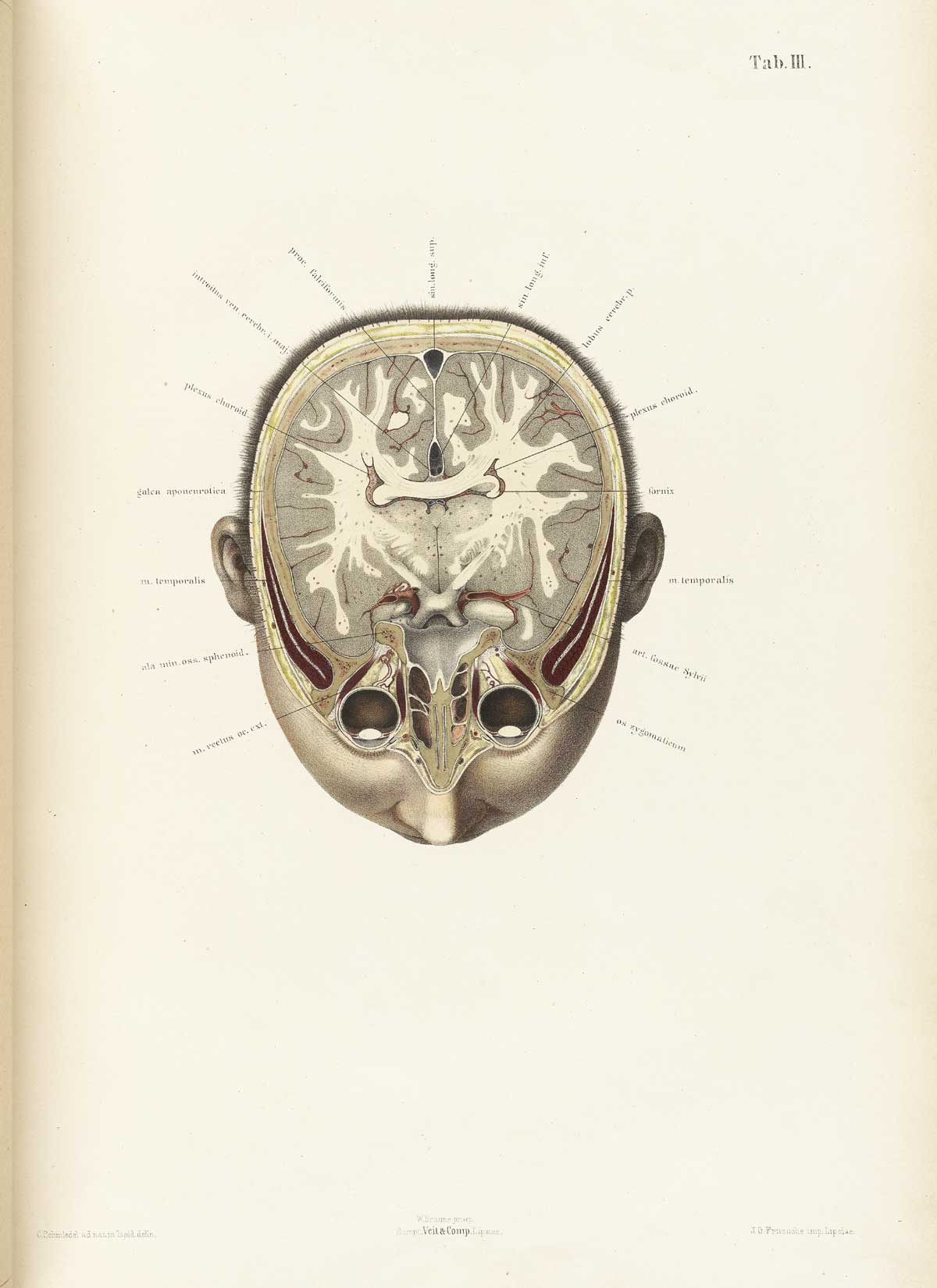
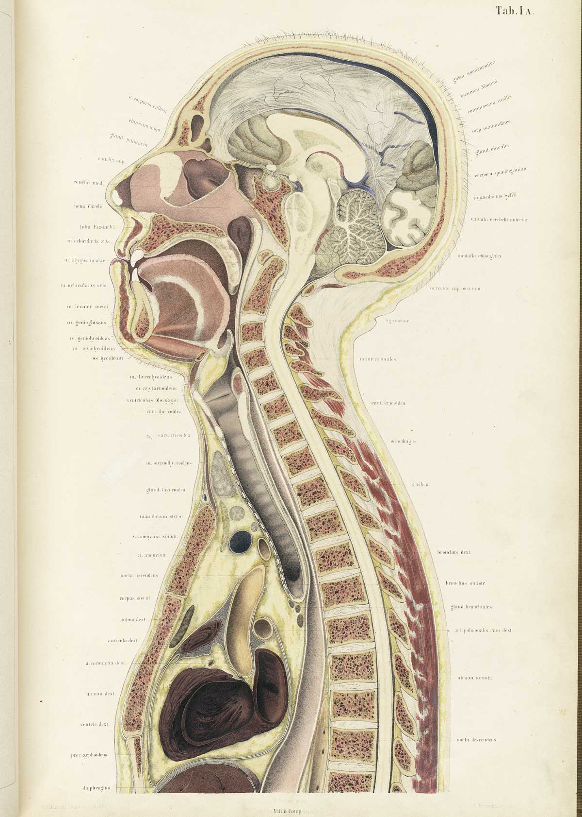
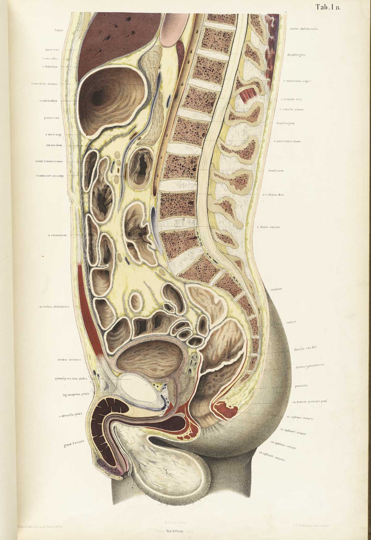
Source [National Library of Medicine]: Braune, Wilhelm. Topographisch-anatomischer Atlas : nach Durchschnitten an gefrornen Cadavern. (Leipzig: Verlag von Veit & Comp., 1867-1872). And Supplement: Braune, Wilhelm. Die Lage des Uterus und Foetus am Ende der Schwangerschaft. (Leipzig: Verlag von Viet & Comp. , 1872).
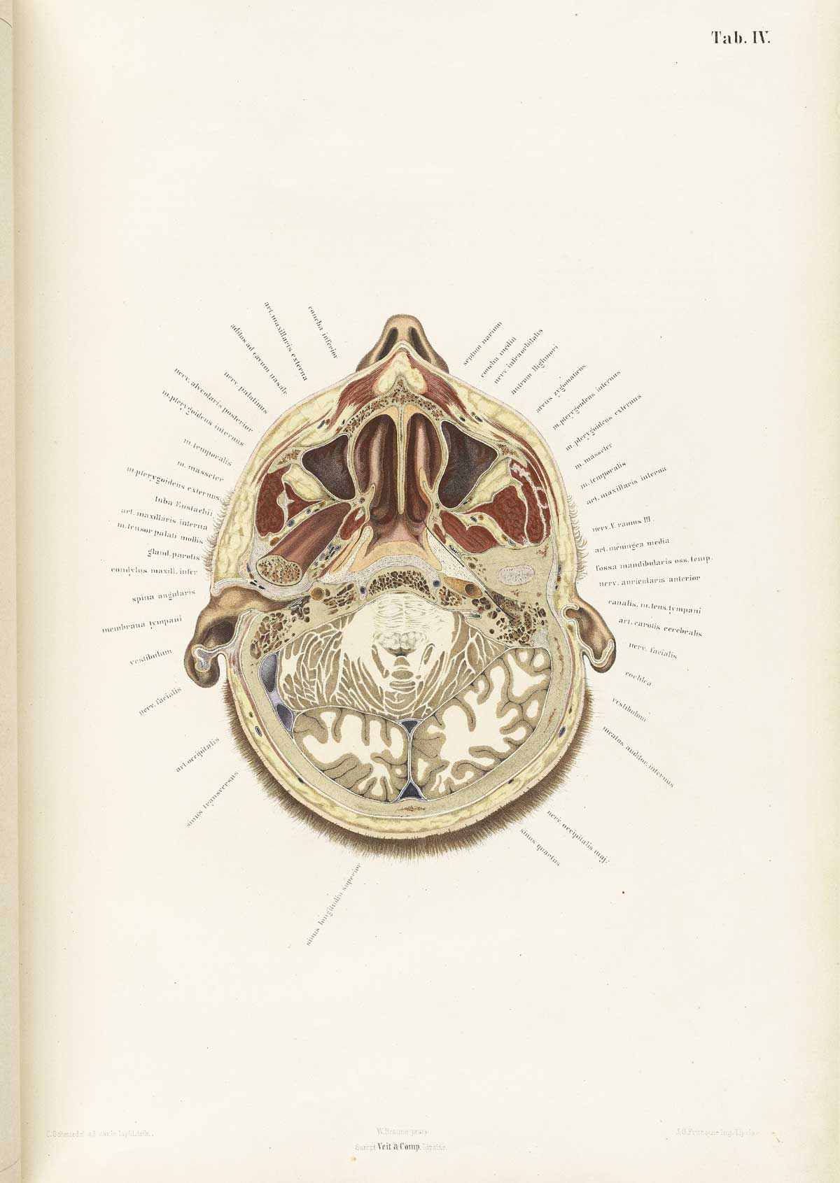
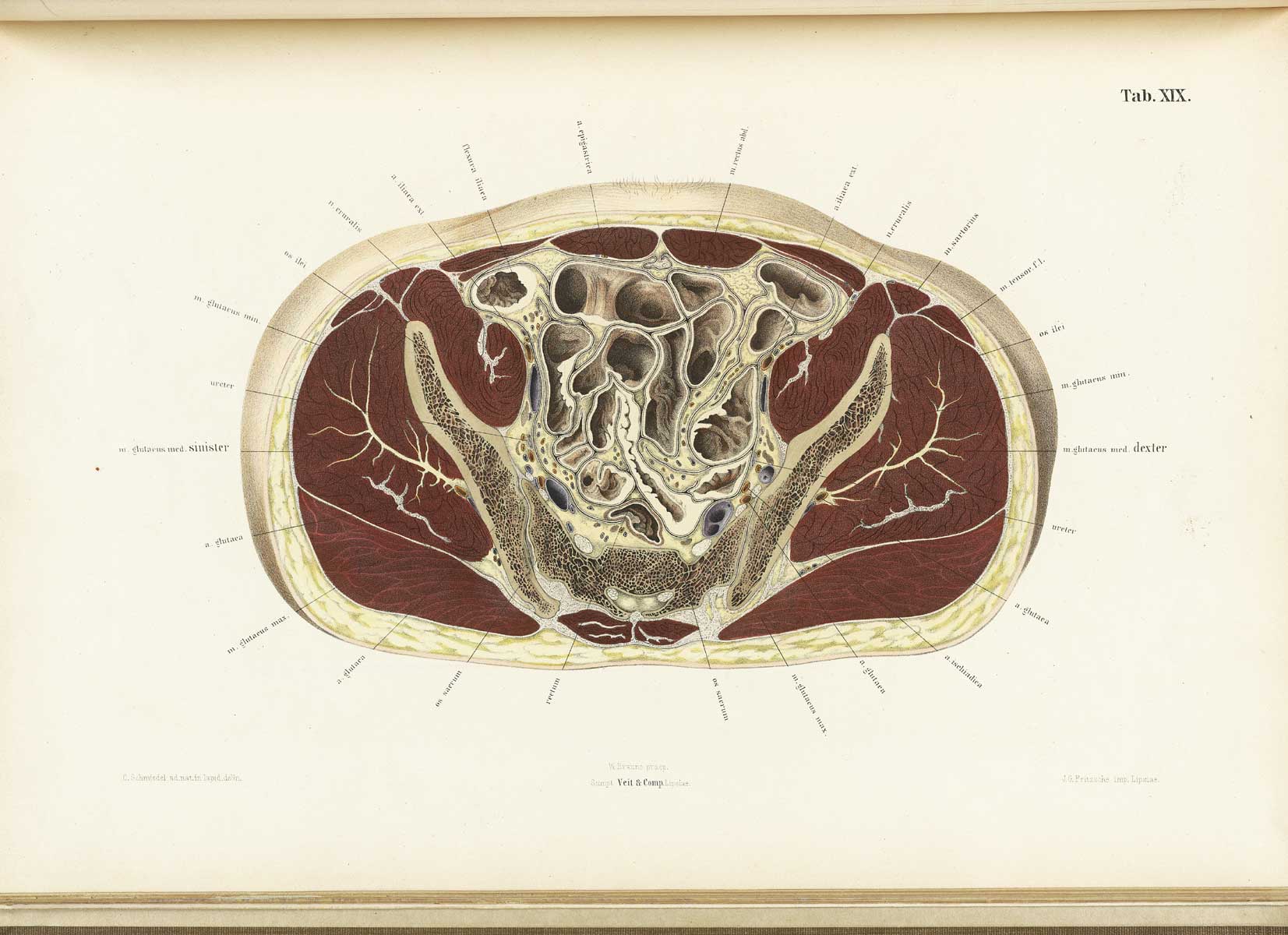
This extraordinary work is part of an anatomical flap book by Wilhelm Braune (1831-1892), a professor of anatomy at the University of Leipzig. The image above is a cross-section of the cadaver that was frozen and thinly cut in the similar vein (sorry!) as the more modern Adam & Eve of the Visible Human Project
 and which consists of over 30 color lithographs of frozen cross sections of human anatomy. The male images from the series are described by Braune:
“[they are] taken from the body of a powerful, well-built, perfectly normal man, aged 21, who had hanged himself. The organs exhibited no pathological abnormalities. The body, which was brought unfrozen, was placed on a horizontal board … In this position, the subject lay untouched in the open air, and at the temperature of about 50 degrees F., for fourteen days. At the end of this time the process of freezing was commenced and completed …”
The National Library of Medicine then continues with the description for preparation of the specimens:
“After sections were cut, thin paper was placed over them and tracings were made of the anatomical features. Similar techniques were used to create the supplement, Die Lage des Uterus und Foetus am Ende der Schwangerschaft, which features large folded plates. Tables B and C of the Supplement include large flaps showing different views of the lower half of the body, and scans were made of each flap along with the shared upper half (i.e. Table B (view 1), Table B (view 2), etc.).”

Notes:
Thw work was published in English as: Braune, W. An atlas of topographical anatomy after plane sections of frozen bodies. Trans. by Edward Bellamy. (Philadelphia: Lindsay and Blakiston, 1877)