A Daily History of Holes, Dots, Lines, Science, History, Math, Physics, Art, the Unintentional Absurd, Architecture, Maps, Data Visualization, Blank and Missing Things, and so on. |1.6 million words, 7500 images, 4.9 million hits| Press & appearances in The Times, Le Figaro, Mensa, The Economist, The Guardian, Discovery News, Slate, Le Monde, Sci American Blogs, Le Pont, and many other places… 5000+ total posts since 2008.

Category: Medicine, History of

  • Questionable Quidity and the Road to Achieving a Faster and More Productive Smoking-Related Disease

    JF Ptak Science Books  Quick Post

    Cigarette manufacturers–like anyone other entity in business–want their clients to consume as much product as possible.  Or buy their goods as often as possible.  With that in mind I’m a little surprised that the Big Five never adopted any of the ideas shown below, all helping the smoker to get at their next cigarette as quickly as possible. For example, in the event hat there were no ashtrays around, several people patented their notions for in-the-cigarette-pack ashtrays, so that no matter where a person was, they could always clicks their ash and stow their butts back in the pack.   Of course, I’m sure that the Makers determined almost instantly that this concern affects perhaps an immeasurable percentage of smokers; if someone wanted (read “needed”) a cig, and there was no ash tray, they can just, well, smoke their cigarette and not use an ash tray. Or go outside.  OR something.  Or perhaps the manufacturers recognized that they could actually sell the idea to some of their smokers, but then realized instantly that carrying around ciggie waste is not attractive and perhaps a stinking mess.  Or they recognized that–like Lee Iaccoca fighting seat belts because it made cars look “unsafe”–having  place to deposit cigarette waste right there with the cigarettes might send a message to the consumer that their product is messy and in the end very unattractive.  Best leave the unattractive bits in a tray somewhere else.  Or of course share the end product of smoking enjoyment with others by leaving it on the ground.

    Then of course to remind consumers how useful the process of smoking is, there were other inventions that combined the products of this act with some other highly useful enterprise, like this cigarette lighter/pencil sharpener with what might have been a whistle attachment:


    And if you found yourself needing/wanting a smoke but didn’t have a free hand, there was always the following device, which allowed the consumer to smoke their stuff through a tube:

    Speaking to the humility of smokers was this device that allowed the user to extinguish their smoking product automatically, no matter what the size of the burning member, reminding the user that if they wanted to actually stop, they could:


    Again, another extension of the art of cigarette smoking and how useful smoking implements can be–the cigarette lighter with built-in flashlight:

    Cigarette lighter with built-in extinguisher:


    Pen with built-in cigarette lighter (1977):

    And another example of a very complicated instrument for extinguishing a cigarette, sending a message with I-don’t-know-what undertones:

    These are just some of the many glorious appendages to smoking and making it easier–many more can be found at the U.S. Patent Office, like all of the examples above. 

  • Disturbing and Disturbed Obstetrical Devices: Straps, Belts and Iron Bits

    JF Ptak Science Books   Post 1501

    Where does a device like the one, below, belong, exactly?  Where in the long rolls of intellectual history should it be found?

    E.A. Agerton is actually Ella C. Agerton–it was easy for me to assume that the person Agerton was a man, because to my experience when I find such devices in the patent office archives they are usually made by men. What Ms. Agerton was attempting with this device is something I don’t understand, though she does make the following statement in her Patent Office record:


    Here’s another, earlier version of this beast, displayed in practical use.  I have to say that the drawing of the woman’s face is deeply disturbed:

    I’ve also found quite by chance that the drawings of the faces of the women drawn as “patients” of these devices can be very unsettling, hardly faces at all, even though the representation of the rest of the body is functional.  For example, the face of the woman below looks as though it is part of the furniture/device:

    The drawings of faces can be so bad that I wonder if anyone can actually be that bad of an artist without trying to be.

    None if this even reads comfortably; I don’t know what it would have been like to been trussed up in such a thing. 

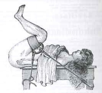
    Blogonbstetrics271 This leads to another uncomfortable image.  At first glance I thought it was part of a delivery procedure, but it turns out that this was the rigging for a woman undergoing a gynecological examination.  The woodcut appears in the first volume of Sammelband von 7 Kleinschriften zu gynakologischen Spezialthemen, 1858-1901, and more exactly in the contribution by Ludwig Neugebauer on an examination with a “speculum bivale l’ecarteur vaginal a trios branches…”  published in 1884.  I know that the prissy prejudices brought about with the coming of Queen Victoria in 1837 still had another 17 years to go at this point, which could explain why, perhaps, a doctor would want an aid in helping a woman maintain an examination position without him having to touch his patient, but, well, just look at the massiveness of the restraints!  Also, the woman’s arms are *inside* the ropes—not down, not holding the ropes, but inside the binding, restrained.  Either way, it isn’t good, treating the woman—in either case of pregnancy or obstetric examination—as the inferior vessel.
    This is also part of a long history of the subjugation of women—the medical aspects of that indoctrination were perhaps somewhat more heinous than the social/legal/ethical/pedagogical/moral constituents.  This illustration, even coming at the end of the Victorian era and approaching the dawn of encroaching bits of equality for (white) women, is a fair example of their subordinate status.

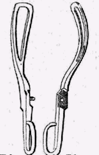
    I just happened upon this picture during the course of my business day—a random image compiling these thoughts.  If I were to try and choose another example of the medical aspect of the subjugation of woman I think that I would choose the story of the Chamberlen family.  For several generations, beginning around 1680, this family of medical doctors practiced a secret—their secret—in delivering breeched babies.  Breeched babies are those who were born feet first, which was usually a death sentence for both the child and the mother.  Patriarch William Chamberlen invented a device that would greatly aid in delivering babies in this position—the forceps.  And evenForceps though everyone today knows what these things are, the Chamberlens kept them a secret from the inquisitive medical world for more than a century.  Their instrument was shrouded—literally!—in secret(s), used under cover, with ceremony, conveyed to the delivery area in a box and brought out with no witnesses.  The Chamberlens felt their invention to be their own, for their own particular use and monetary gain, and the rest of the world be damned, and at least three generations (grandfather, father, son) kept the family secret. If their family could not be in charge of such a delivery then there would be no use of their extraordinary invention.  This of course would save their income from being chipped away by other physicians or midwives or whatever competent person could be found to utilize the forceps—their economic gain coming at the cost of (savable but imperiled) pregnant women who would either pay the Chamberlens for aid in their breeched births or, well, probably die.     To what degree this revolting behavior damaged other people can not be calculated. I’m not even sure about how to translate this into something more recognizable—would it be like Robert Koch keeping his isolation of Bacillus anthracis (1877), or  tuberculosis bacillus (1882) a secret?  Or Wilhelm Roentgen shrouding his discovery in secrecy somehow? (I know, I know, this is impossible.)  Or isolating a secret, proprietary cure for cancer?
    There is a large part of me that believes that if the Chamerlens had figured out something like this that affected only men, a silver bullet for something or other, that their secret wouldn’t stayed a secret for 100 hours, let alone 100 years. 


  • Electro-LUXurious 4: the Curative Electric Chair, Complete with Apparatus for the Treatment of the “Depending Organ”, 1891

    JF Ptak Science Books   Quick Post

    This is the fourth in a new series of posts on interesting, early applications of electricity, most of which are taken from the archives of the U.S. Patent Office.

    J(osephus) C. Chambers, the patentee of the apparatus below, was the owner of Chambers’ National Lightning Protection Company (Newport, Kentucky), and held a number of patents relating to lightning rods, electrical insulators, a telephone apparatus (1881), and a number of associated bits of techno. I’m not exactly sure what this device, below, did, except that it seems like a low-voltage quack-medical electric chair.

    The first patent drawing, below, shows a view of the chair and at front, looking backwards towards the  “ornamented” wooden cabinet that contained the guts of the machine, with the seat (which would have a metallic, conductive surface) removed.  You would place your hands on the two capital “H” pads that seem to be just below the two drawers; your feet would go on the two “h” pads at bottom. 

    The drawing below references how the various swinging arms and such can be maneuvered to reach any part of the body.  Mind you now the inventor doesn’t actually state what this electrical treatment “does”, only how you apply it and the technical stuff behind the machine. Also ir is not a solitary experience–there would be another person operating the controls on the top of the wooden case.

    I wondered about the cup (“J”), and thought, well, maybe that’s not what I think it might be for…but it is, waiting there to treat “the depending organ”.


    The U.S.  Patent Office report reads as follows:

    United States Patent Office.

    JOSEPHUS C. CHAMBERS, OP DETROIT, MICHIGAN.
    ELECTRICAL MEDICAL APPARATUS.

    SPECIFICATION forming part of Letters Patent No. 445,636, dated February 3, 1891.

    Application filed April 21,1890, Serial So, 348,911, (So model,)

    To all whom it may concern:


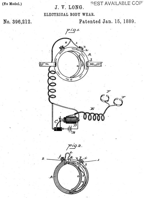
  • Electro-LUXurious 3: Anti-erection “Body Wear” 1889

    JF Ptak Science Books  Quick Post

    Electrical Anti-Erection “Body Wear”  1889

    This is the third in a new series of posts on interesting, early applications of electricity, most of which are taken from the archives of the U.S. Patent Office.  Unlike the general post in this blog, many of the Electro-LUXurious post images and documentation can be presented without commentary. 

    Patent anti erection


    [Source: Google Patents, http://www.google.com/patents/US396212]


  • An Alphabet of Anatomical Emotions and Feelings–Installment 1

    JF Ptak Science Books   Post 1488

    In a developing thread on oddly-represented alphabets on this blog, this one is a little harder to fill than it seems.  The rules are that all images must be some level of dissection (with the exception of the anatomical Durer), and not a simple artistic study of emotion, and that the images be at least 200 years old.  Part I of this alphabet of emotion and feelings includes awe, burdensome, contempt, disappointment, empathetic, fear, guilt, hope, intrigue, joy, lonesome, majesty, pride, resentment, surprise, trust, vulnerability and wonder.

    Awe

    Tabulae Anatomicae... by Giulio Casserio and Odoardo Fialetti

    Giulio Casserio (ca. 1552-1616, anatomist) and Odoardo Fialetti (artist), Tabulae Anatomicae, published in Venice in 1627.

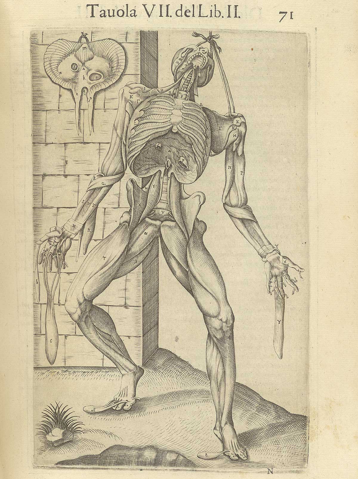
    Burdensome

    http://www.nlm.nih.gov/exhibition/historicalanatomies/Images/1200_pixels/valverde_p71.jpg

    Juan Valverde de Amusci (ca. 1525-ca. 1588), Anatomia del corpo humano, printed in Rome in 1560. 


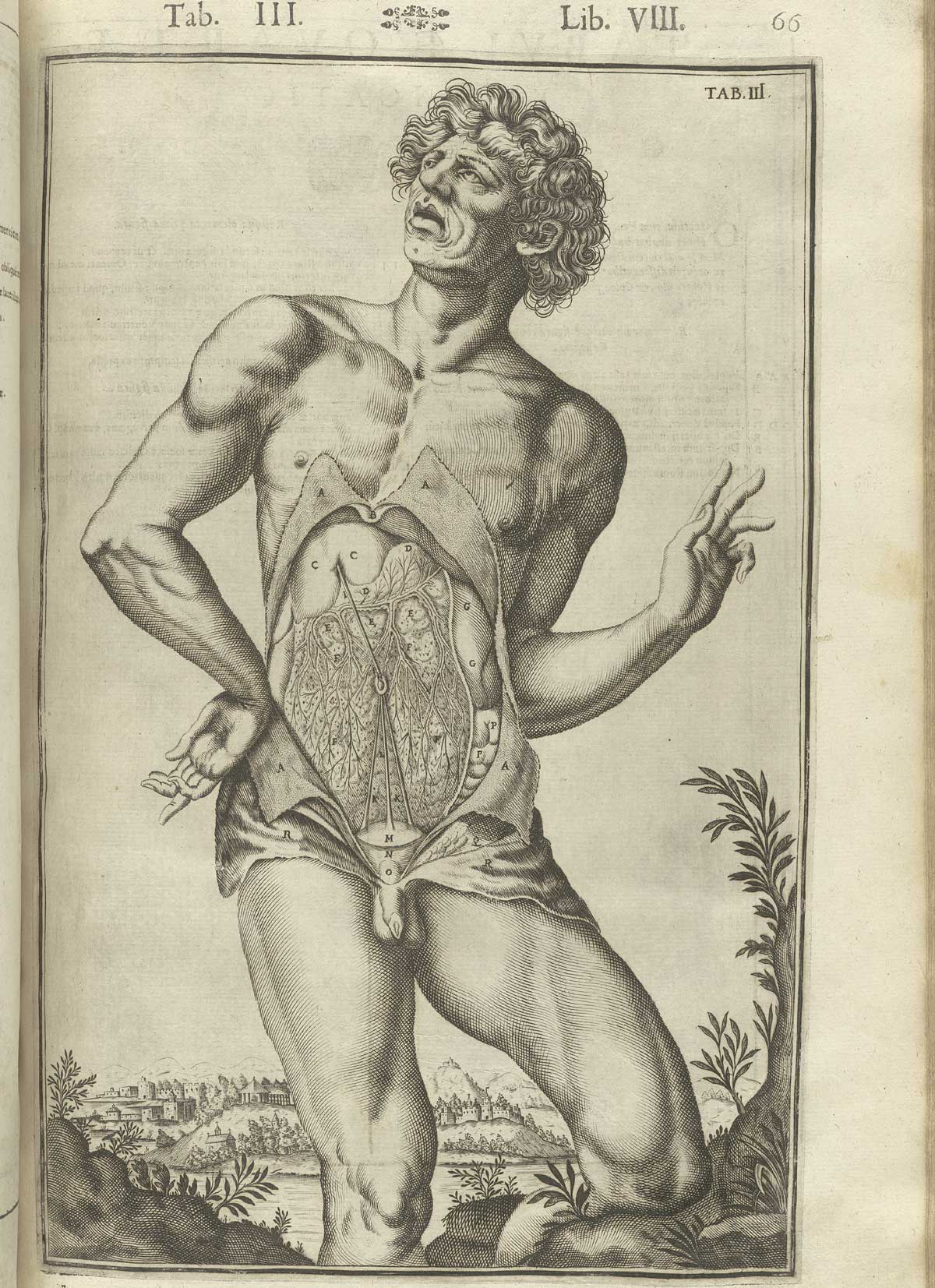
    Contempt

    Casseri_p66  dandy

     Giulio Casserio (ca. 1552-1616, anatomist) and Odoardo Fialetti (artist), Tabulae Anatomicae, published in Venice in 1627

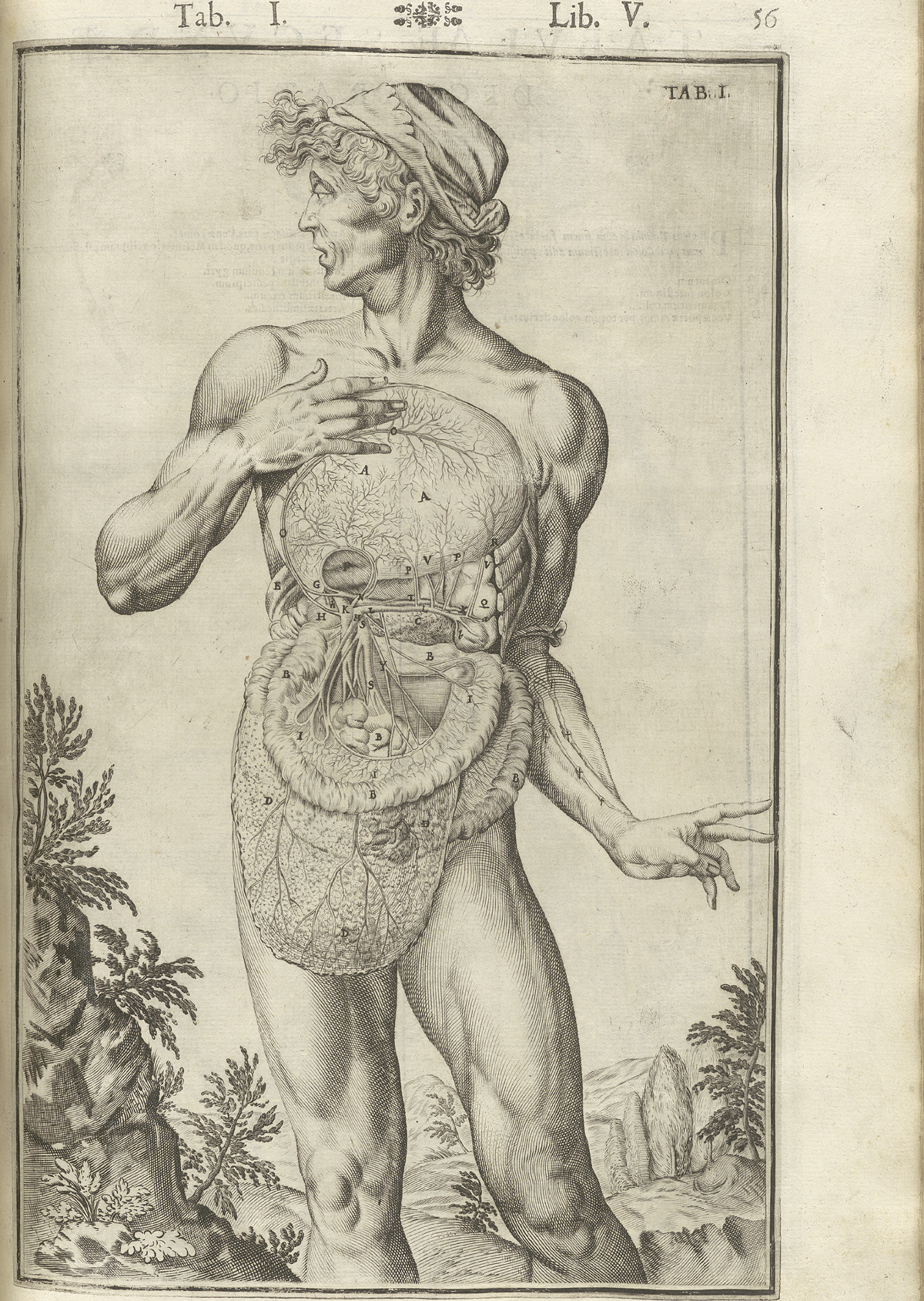
    Disappointment 

    Casseri_p56 surprise

    Giulio Casserio (ca. 1552-1616, anatomist) and Odoardo Fialetti (artist), Tabulae Anatomicae, published in Venice in 1627.

    Empathetic

    Missing skin c
    Juan Valverde de Amusci (ca. 1525-ca. 1588), Anatomia del corpo humano, printed in Rome in 1560.

    Fear

    Gamelin1_t02 Jacques Gamelin (1736-1803), Nouveau receuil d’ostéologie et de myologie, published in Toulouse in 1779.

    Guilt

    Gamelin1_t11
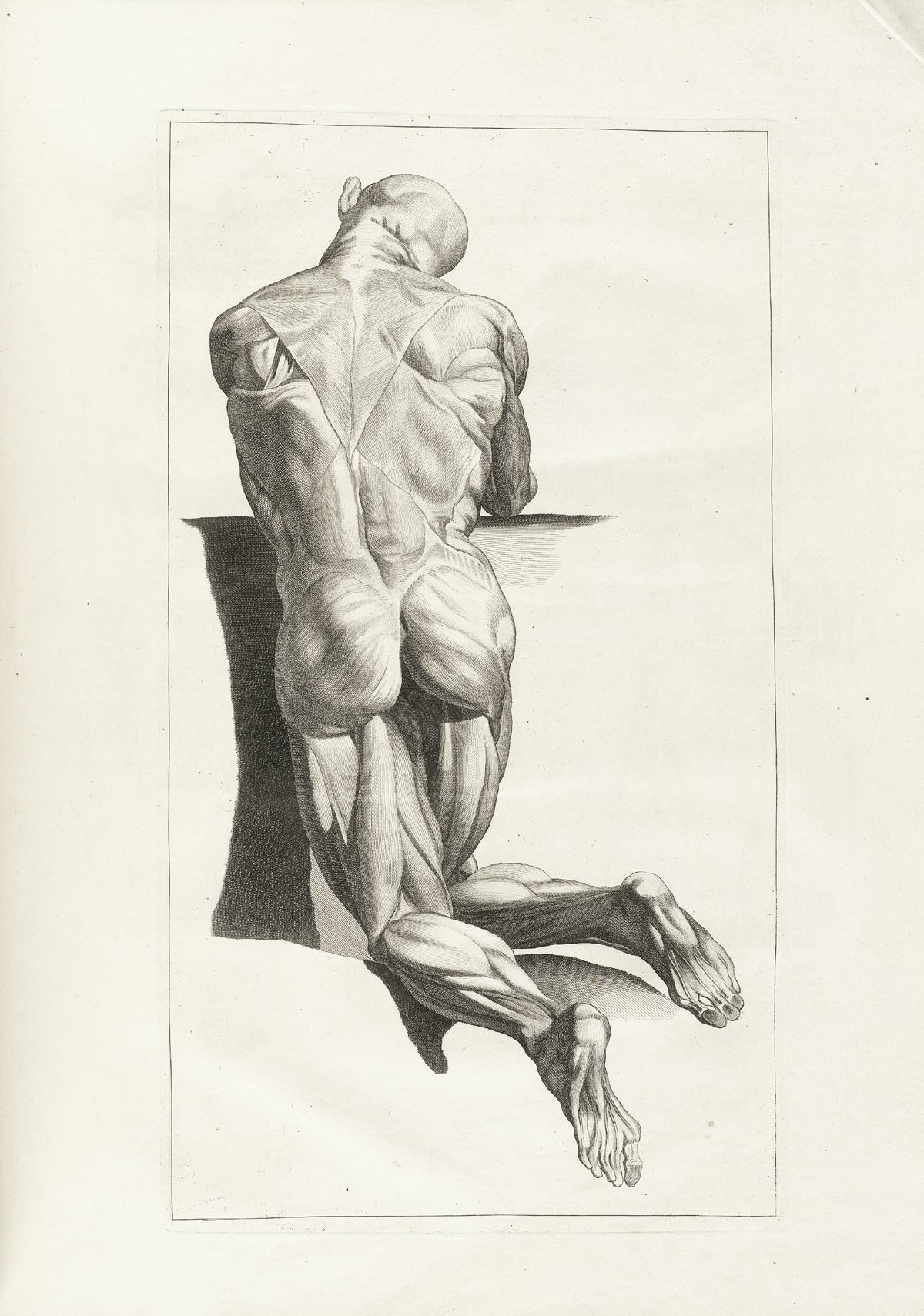
    Jacques Gamelin (1736-1803), Nouveau receuil d’ostéologie et de myologie, published in Toulouse in 1779.

    Hope

    http://www.nlm.nih.gov/exhibition/historicalanatomies/Images/1200_pixels/gersdorff_p21v.jpg

    Hans von Gersdorff (d. 1529),  Feldtbůch der Wundartzney : newlich getruckt und gebesser, published in Strassburg in 1528.  

    Intrigue

    Casseri_p21 bored

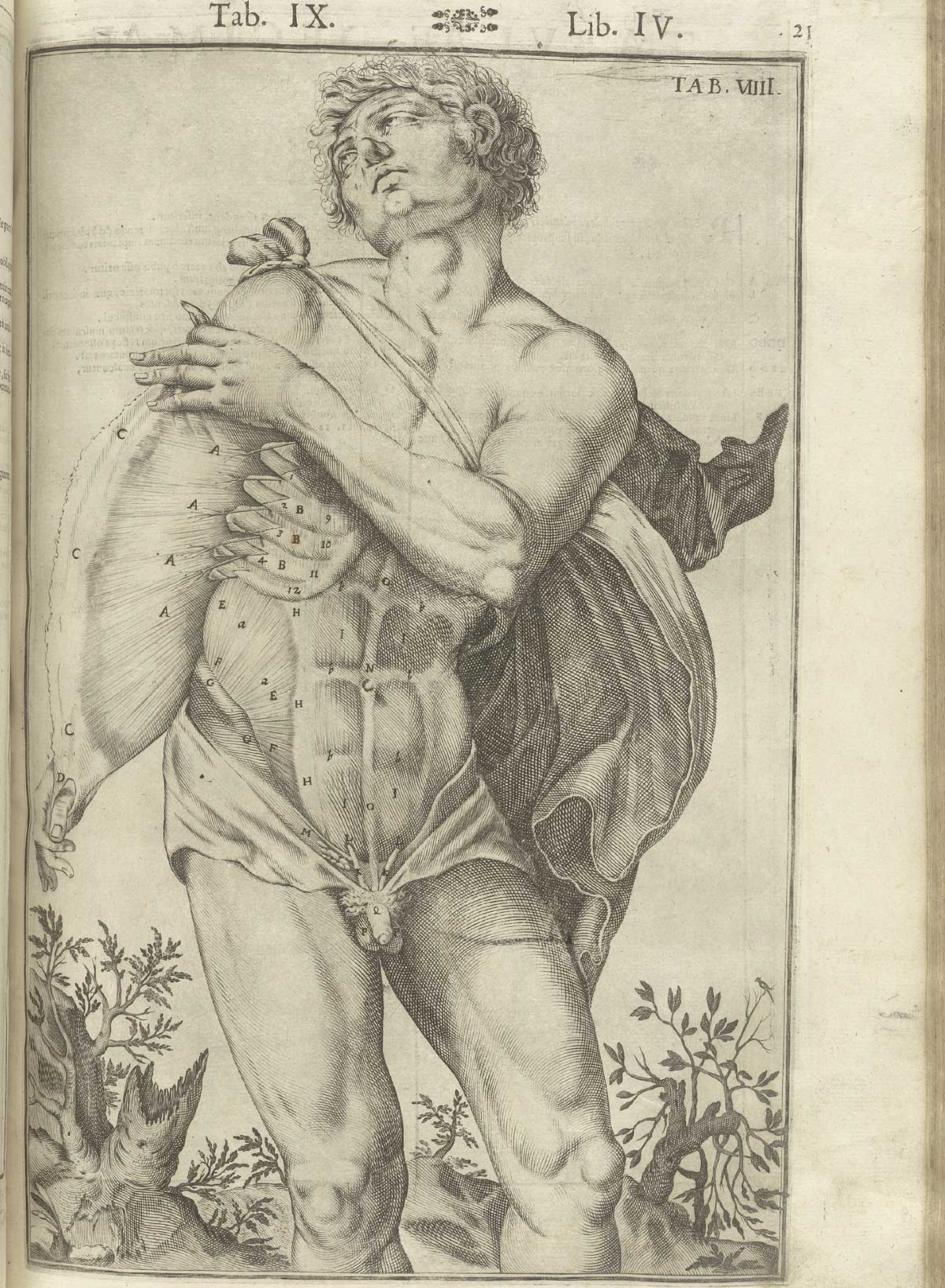
    Giulio Casserio (ca. 1552-1616, anatomist) and Odoardo Fialetti (artist), Tabulae Anatomicae, published in Venice in 1627

    Joy

      Anatomy john brown dancing

    Lonesome

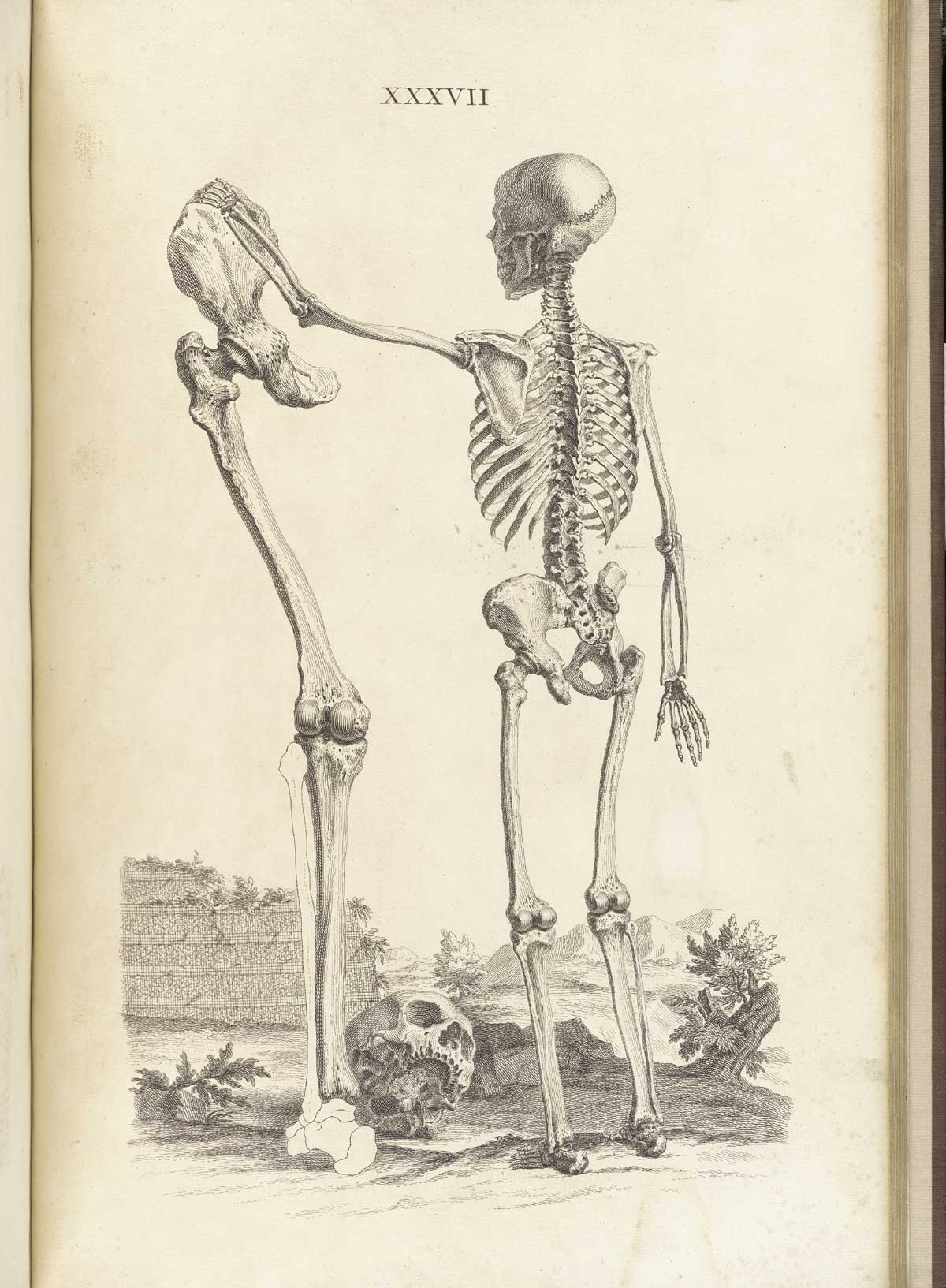
    http://www.nlm.nih.gov/exhibition/historicalanatomies/Images/1200_pixels/cheselden_t37.jpg

    William Cheselden (1688-1752), Osteographia, or the Anatomy of Bones, published in London in 1733.

    Majesterial

    http://www.nlm.nih.gov/exhibition/historicalanatomies/Images/1200_pixels/Eustachi_t39.jpg

    Bartholomeo Eustachi (d. 1574), from Tabulae anatomicae, printed in Rome in 1783.

    Pride


    Anatomy performers

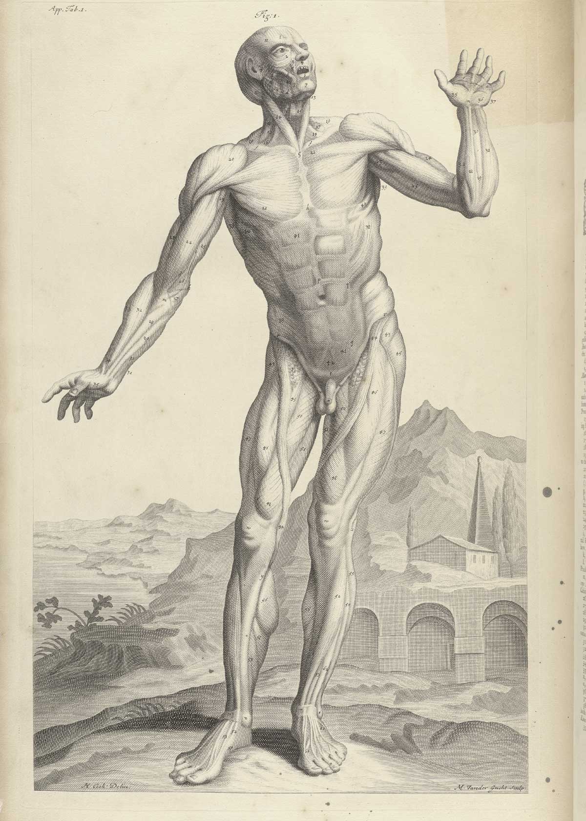
    In the category of “Holy Crap!” anatomical illustration surely Pietro Berrettini da Cortona (1596-1669) must be among its patron saints.  His images are incredible, yelling and screaming for attention as they demand their space onthe printed page.  Further, the man actually accomplished all o fthis in the early 17th century, his work no tbeing published for another 130 years or so. [Source:  Tabulae anatomicae..., Rome 1741.]

    Resentment

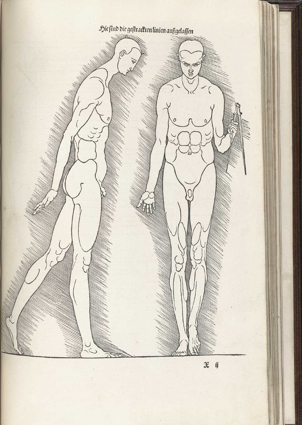
    http://www.nlm.nih.gov/exhibition/historicalanatomies/Images/1200_pixels/durer_pg-233.jpg

    Albrecht Durer (1471-1528), Vier Bücher von menschlicher Proportion, published in Nuremberg in 1528.


    Surprise
    http://www.nlm.nih.gov/exhibition/historicalanatomies/Images/1200_pixels/cowper_t01.jpg

    William Cowper, The anatomy of humane bodies, printed in Oxford in 1698.


    Trust


    http://www.nlm.nih.gov/exhibition/historicalanatomies/Images/1200_pixels/cheselden_t36.jpg

    William Cheselden (1688-1752), Osteographia, or the Anatomy of Bones, published in London in 1733.

    Vulnerable

    Hundt_p119

    Magnus Hundt (1449-1519),  Antropologium de hominis dignitate…, printed in Leipzig in 1501 by Wolfgang Stoeckel. 

    Wonder

    http://www.nlm.nih.gov/exhibition/historicalanatomies/Images/1200_pixels/Estienne_p043.jpg

    Charles Estienne (ca. 1504-1564), De dissectione partium corporis humani libri tres, published in Paris and printed by Simon Colinaeus in 1545.


  • Species of Explosions: Human Anatomy, Explained

    JF Ptak Science Books   Post 1479

    Anatomia universale... by Paolo Mascagni and Antonio Serantoni

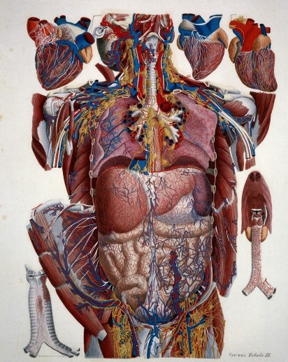
    Species of explosions come in all sorts of shapes and sizes, and basically release some sort of energy (chemical, nuclear, mechanical and so on), but in the cases we’ll look at here they are none-of-the-above.  The energy released in these explosions are in the form of potential, a knowledge-driven, controlled and reversibly ordered demolition, a deconstruction in slow motion.  The medical “explosion view” for the study of anatomy is a remarkable thing, especially when you considered not only the pre-Cat and pre-MRI times, but also the pre-Xray (1895) days of imaging the inside of the body–the images not only show you the bits and pieces alone but also in relation to the other bits around them.  The body becomes an archaeological dig, each level preserved so that a viewer could easily see the dependencies and relations between one thing and another. 

    Take for example Paolo Mascagni’s (1755-1815) beautiful Anatomia universale (an example seen above), which was printed in Florence in 1833 and has the look and sensibility of something much later.  Mascagni is able to achieve this depth not only from the exploded view, but also for the (somewhat exaggerated) coloring setting the specimen on top of a blank background the combination of the color and the background giving the whole thing a 3-D-ish feel–and if not that, then certainly a living image in which there is some fair amount of depth. 

    Anatomy exploded
    Another version of the exploded view which is also quite effective from the same work by Mascagni is his “exploded thorax”, showing a more limited display of the more closely-aligned organs. 

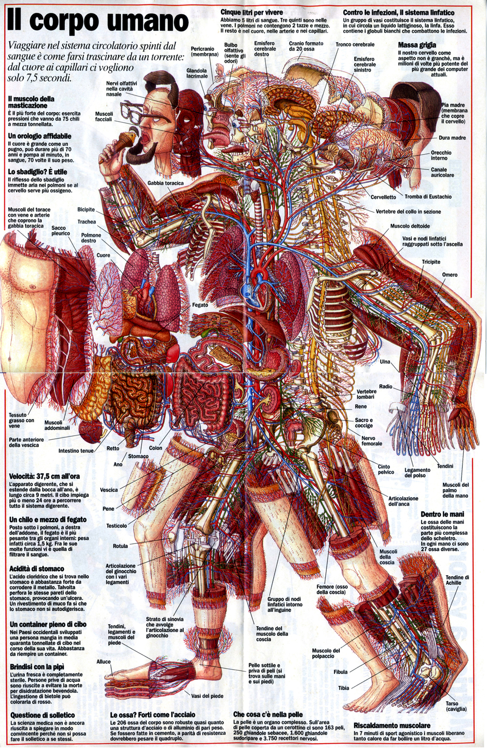
    When these views appear today–which is generally much more uncommon given the other technical ways of envisioning multiple layers of complex associated items–they have more of a sense of the antiquarian, retro feel to them, though they are no less useful for it.  As a matter of fact  I still prefer these views for understanding technical and engineering  and architectural complexes than anything else. And the modern master of this genre for me is Stephen Beisty, who performs his magic mostly in the tech and engineering fields, but who occasionally dips into the messy wet stuff of biology, as we can see in this fabulous exploded view:

    Anatom exploded beisty

    There are of course many other examples of (drawn) exploded views in the history of anatomical illustration, but I think these will do for now as good examples of the art.


  • Things-out-of-Place Department: Books in Anatomies and Bridges in Impressionist Paintings

    JF Ptak Science Books   Post 1478

    I’ve been making a few entries over the last few days on antiquarian anatomy books, and wonder now about books in anatomies. I’m not certain what the appeal is, but it has a strange and immediate affinity for me to bridges in pre- and early Impressionism, a direct line to these bridges and to the other technological and engineering entities that were depicted in these canvases.  I’m not sure what the connection is to the two outside of the obvious verb-y part of “bridge”, but there seems to be an innate connection between the two, for me.  Somehow the book shouldn’t be of interest to the lifeless and dissected cadaver, but it is, and images of the two together turn up here and there in the history of anatomical illustration.  As much as the anatomist and artist work together to try to remove their subject from the confines of a book so that their work is freer and more accessible to the reader, to remove the illustrations from the pages of the book in which they appear, they do sometimes employ the image of the book to make a more prosaic and allegorical point.And as the object of design and wonder and beautiful geometry, one might think of the bridge as a object in Impressionist paintings from that movement’s earliest periods would be not quite the sort of thing that would be the subject of as many paintings as they were.  The unnatural angles of their supplementary parts  seem to make them not quite the fit for the early Impressionists.  But they were, and probably so for their 90-degree assaults on the natural landscape. 

    Perhaps what I think of the aesthetics of the treatment of bridges during the Renaissance, and the adoration of the Rialto and Pone Vecchio and all the rest, just wouldn’t make their way to the Impressionists’ canvases. But of course basically most ot them did paint bridges–Seurat, Cezanne, Van Gogh (a fantastic, small stone-and-wood miniature drawbridge), Derain, Pissarro, Renoir (the Pont-Neuf), Marquet, Sisley and of course and perhaps most famously, Monet (Charing Cross, Giverny, and all the rest)–all painted bridges.  I guess the bridge is just a shorthand for the representation of engineering and technology in general in Impressionism, which to my eye seems quite strong. 

    But getting back to books in anatomies:

    Ontleding des menschelyken lichaams by Govard Bidloo and Gerard de Lairesse

    Govard Bidloo approached the issue of the book in a most unusual fashion, making it appear as though the dissected hand and forearm were emerging from a semi-opened book, the long index finger coming to a pointing pose, suggesting perhaps that the viewer take heed of what is contained in the work. The image appers in Bidloo’s Ontleding des menschelyken lichaams (or Anatomia Humani Corporis) and was printed in Amsterdam (at what may have been the pinnacle decade of Dutch book publishing) in 1690; the book was illustrated by Gerard de Lairesse (1640-1711), a leading light in the Golden Age of Dutch painting, and perhaps one of the most famous painters alive there after the death of Rembrandt. There’s all manner of symbolism going on here, but what I like the most is the little crutch under the cadaver’s hand’s thumb. 

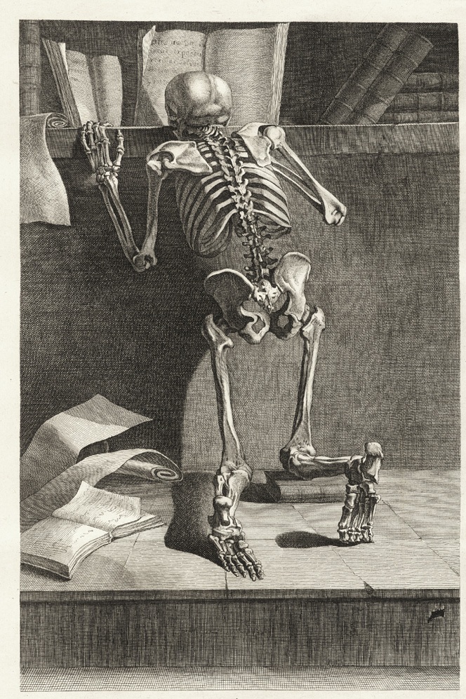
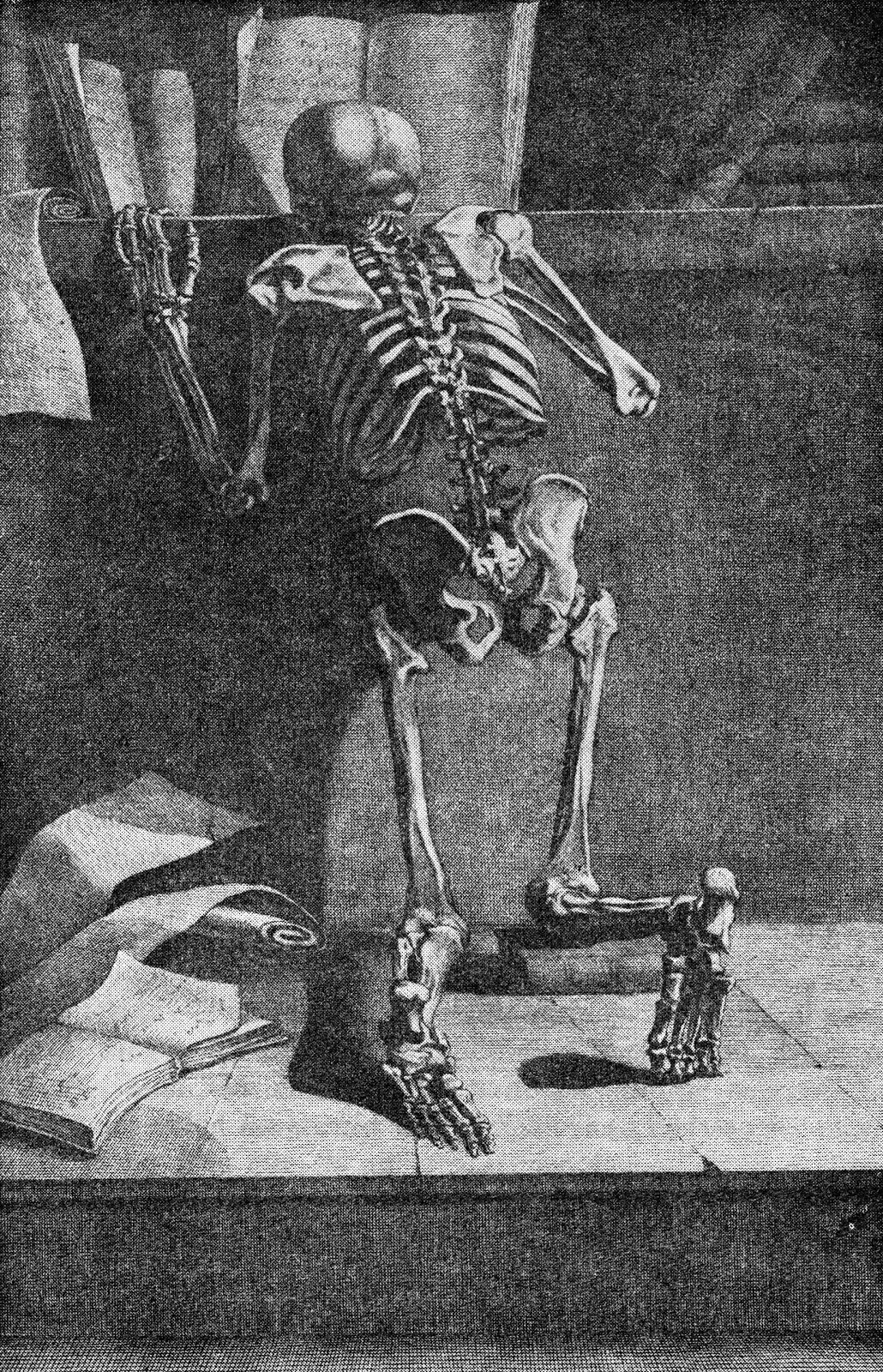
    Jacques Gamelin’s Nouveau reueil d’osteologie et de myologie, dessine d’apres nature (1779)  places skeleton and books togehter again, though this time their can be little doubt about the overall message that the designer and author tried to send to their readers. 

    Int he long run, and I guess in the short run as well, neither the bridge in Impressionism nor the book in anatomies is quite so much out-of-place as they are simply unexpected.

    Note:

    Van Gogh’s bridge at Arles–not an essential image whatsoever for this post–I just like the bridge. Van Gogh painted the bridge four times in oil and once in watercolor, all in a very short time.  (His stay at Arles was about 444 days, during which he made about 200 paintings, being some of his greatest work.

    Vincent Van Gogh Miscellaneous Buildings Technology Modern Age Impressionism Neo-Impressionism


  • Ultra Realism in Anatomical Illustration, 18th Century.

    JF Ptak Science Books   Post 1477

    [In case there are younger readers here, this is a notice that some may consider the following images disturbing.]

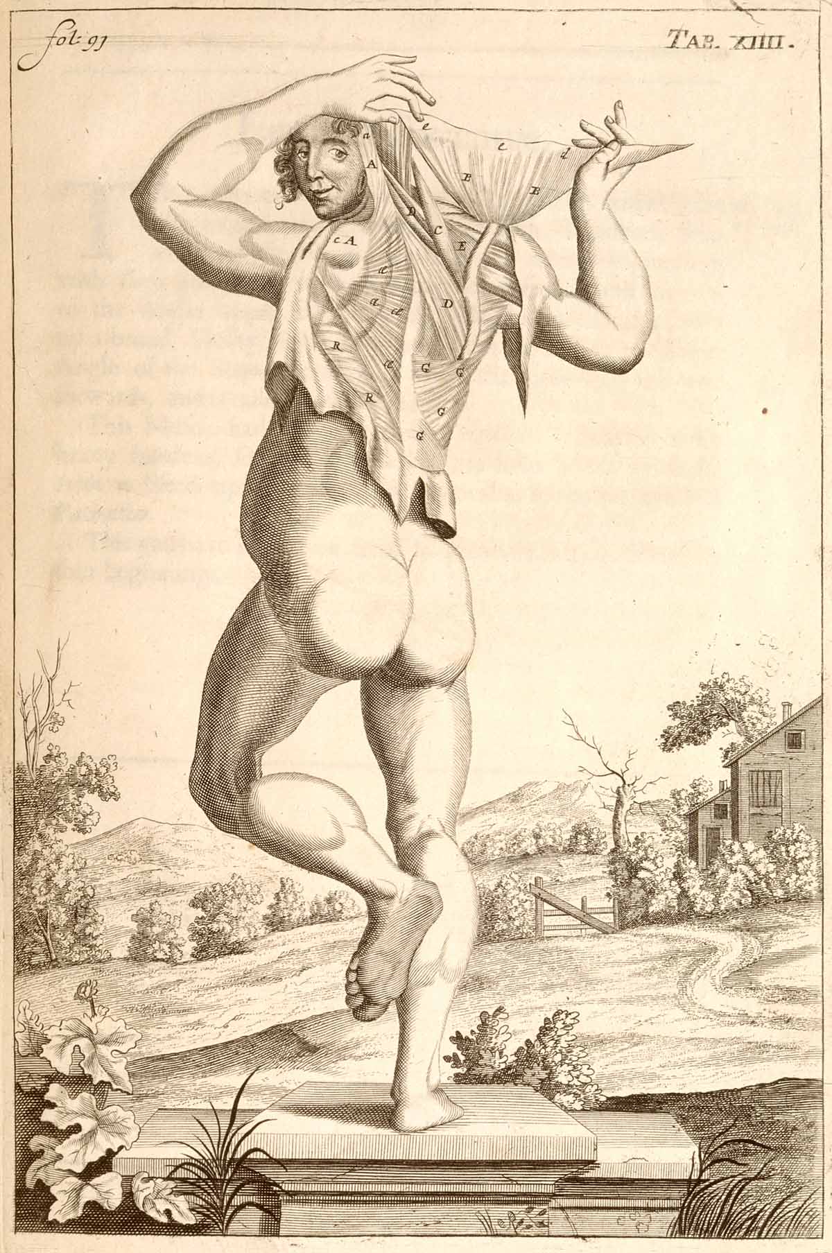
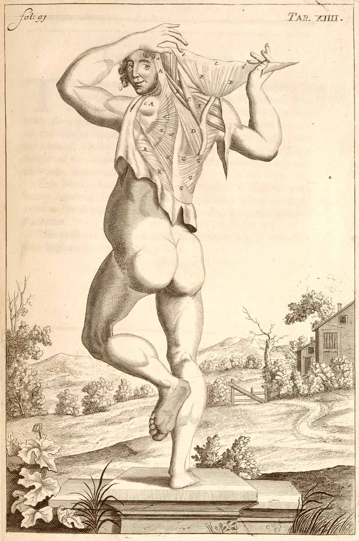
    Anatomy john brown dancing In the antiquarian world of anatomy books dissection and presentation take place on many different levels–there are very straightforward, unadorned displays of various body parts on blank backgrounds and with no attention paid whatsoever to anything beyond the dissection; there are others that place the dissections and cadavers and whatever the subject of dissection might be in prosaic fields, or against a distant cityscape, or in a well-appointed room, or against a generic table, or within some sort of Baroque ornamentation, and so on.  (We’ll be seeing more of these presentations in the next few posts.)  In general though the work of the anatomist is presented in  a basically “finished” state–that is, the dissection is presented in an artistic manner, with a sense of design for the subject material. (Here’s one example of hundreds of possibilities: John Browne’s A Compleat Treatise of the Muscles, as they appear in the Human Body...1681.)

    There are not many examples (before, say 1850) in the history of anatomical display that show the specimens in the stage of display that was almost entirely unadorned, as though we are looking directly at the anatomist’s table.  Such is the case in the extraordinary work of the anatomist Govard Bidloo (1649-1713) and (artist) Gerard de Lairesse (1640-1711) called Ontleding des menschelyken lichaams…, published in Amsterdam in 1690, which is an unremitting attack on realism in the art of anatomy.  There is absolutely no favoritism paid to anything in these images except for truthful display, right down to the nails and pins used to give messy stuff a more-recognizable (to the layman) form. 

    Ontleding des menschelyken lichaams by Govard Bidloo and Gerard de Lairesse
    Ontleding des menschelyken lichaams... by Govard Bidloo and Gérard de Lairesse
    Engravings of the bones, muscles, and joints by John Bell

    Another, later, uncommon example of this realism is found in the 1804 work of John Bell’s Engravings of the Bones, Muscles, and Joints, Illustrating the First Volume of the Anatomy of the Human Body

    Illustrations of dissections by Ellis and Ford

    These images are a world away from the overwhelming majority of works of anatomy produced from the 16th to the 19th centuries; and that Bidloo’s work was so early makes it that much more remarkable. 

    Source note:  all images courtesy of the National Institutes of Health National Library of Medicine, here.


  • Blank and Missing Things Series–Antiquarian Anatomical Skin (Appended)

    JF Ptak Science Books   Post 1476  -part of the Blank, Missing and Empty Things series.

    Carl Ernst Bock (1809-1874) was a physician and anatomist (and son of the anatomist August Carl Bock of Leipzig, 1782-1833), whose many publications include the very popular  Hand-Atlas der Anatomie des Menschen  nebst einem tabellarischen Handbuche der Anatomie.(first printed in 1841).  This is a good example of his reaching out across the social strata to publish something that was accessible to the public-at-large, making him a pioneer in the distribution of medical information.

    I’m sorry to say that in all of this man’s beautiful work I  focused on this superior full-body anatomical–the odd thing here is that the only skin remaining on the body is on the head, where we see a  face and full head of hair.  To my experience this is a very highly unusual visualization.

    Anatomy bock482

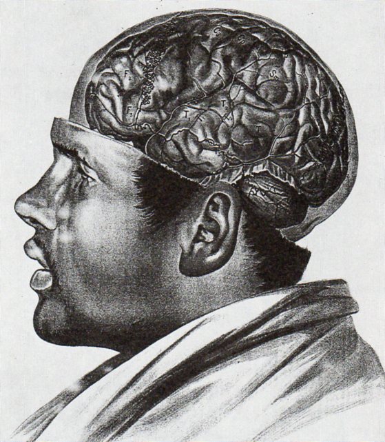
    In an almost-the-opposite approach (which is much more common) Albert Adamkiewwicz (in his Taflen zir Orientierung an der Gehirnoberflaeche des lebenden Menschen...(printed in 1894, in Vienna) offers the following:

    Anatomy adamkiewicz484

    As with the other image above I only have a second generation image of this, and only in black and white–the color original is truly remarkable. 

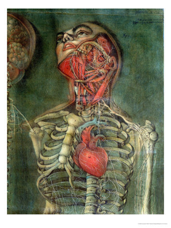
    Another excellent example of an anatomical dissection leaving the face intact occurs several times in the massively interesting work of Jacques Gauthier Dagoty, whose work I remembered but whose name I forgot, making it fairly difficult to find the illustrations.  But here they are, from Anatomie des parties de la generation de l’homme et de le femme, published in Paris, in 1773, with Dagoty (1717-1785)  acting in multiple roles of author/designer/anatomist/painter, who produced these magnificent painterly dissection studies of great detail and warmth.

    Anatomie des parties by Gautier D’Agoty and Duverney

    And this:

    Missing skin b
    There are many other studies in the volume, but few retain the face.

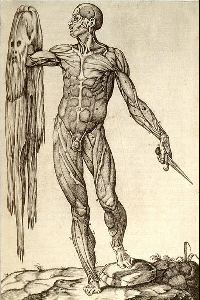
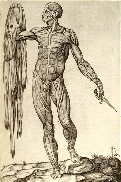
    Then there’s this, the other end of the spectrum, where we see the flayed anatomical specimen and its skin though not in contact with one another, which is a special category indeedy:

    Missing skin c

    This copperplate engraving comes from Juan Valverde de Amusco (Valverde) (1525-1588 or thereabouts), Anatomia del corpo humano… (printed in Rome in 1559), and stands quite apart (though not alone) from the rest of the anatomical oeuvre of the 1550-1750 period.  

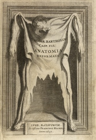
     And let’s not forget Thomas Bartholin’s frontspiece for his Anatomoa, published by F. Hackis in Leyden in 1651, which is similar to the Valverde:

    Anatomy skin bartholoiin


  • Architecture of Insanity

    JF Ptak Science Books   Post 1474

    Insane asylum485

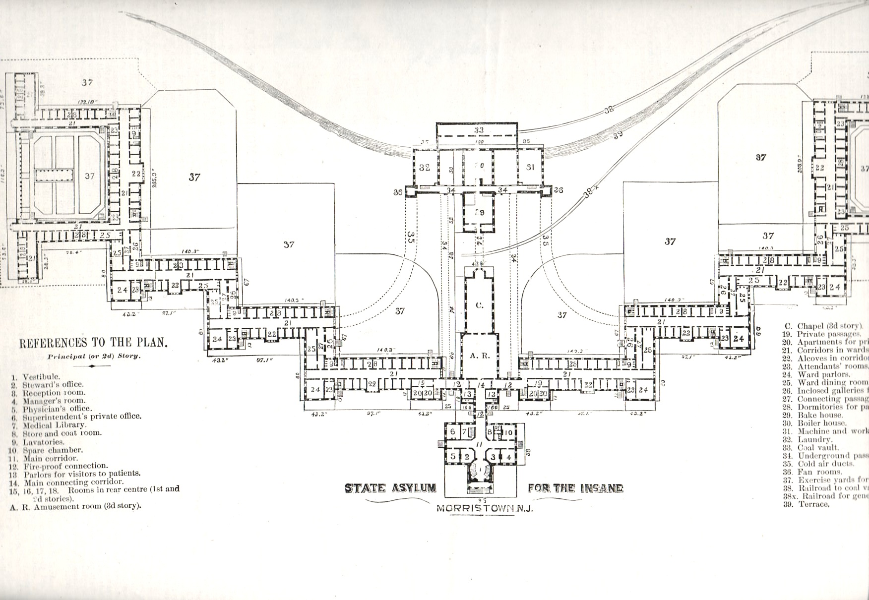
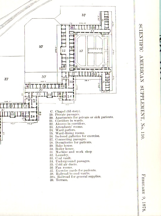
    I was browsing an 1878 issue of The Scientific American, looking for an article on torpedoes, when I came across this stunning architectural plan of the Morristown (New Jersey) State Asylum for the Insane–I recognized its design as something I had seen before, several times, at least.  This design required a lot of land and a lot of bricks–the building was a few stories high and laid out far and wide, built to accommodate 2,000 patients.  The design was intended to allow patients comfortable living space, room for privacy as need be, exercise area and plenty of open yards around the structure for sun and fresh air.  Of course building like this were quickly filled and filled some more, requiring the space for rehabilitation to be used for stockpiling other patients until more appropriate situations arose, which they mostly seemed not to.

    Insane asylum486

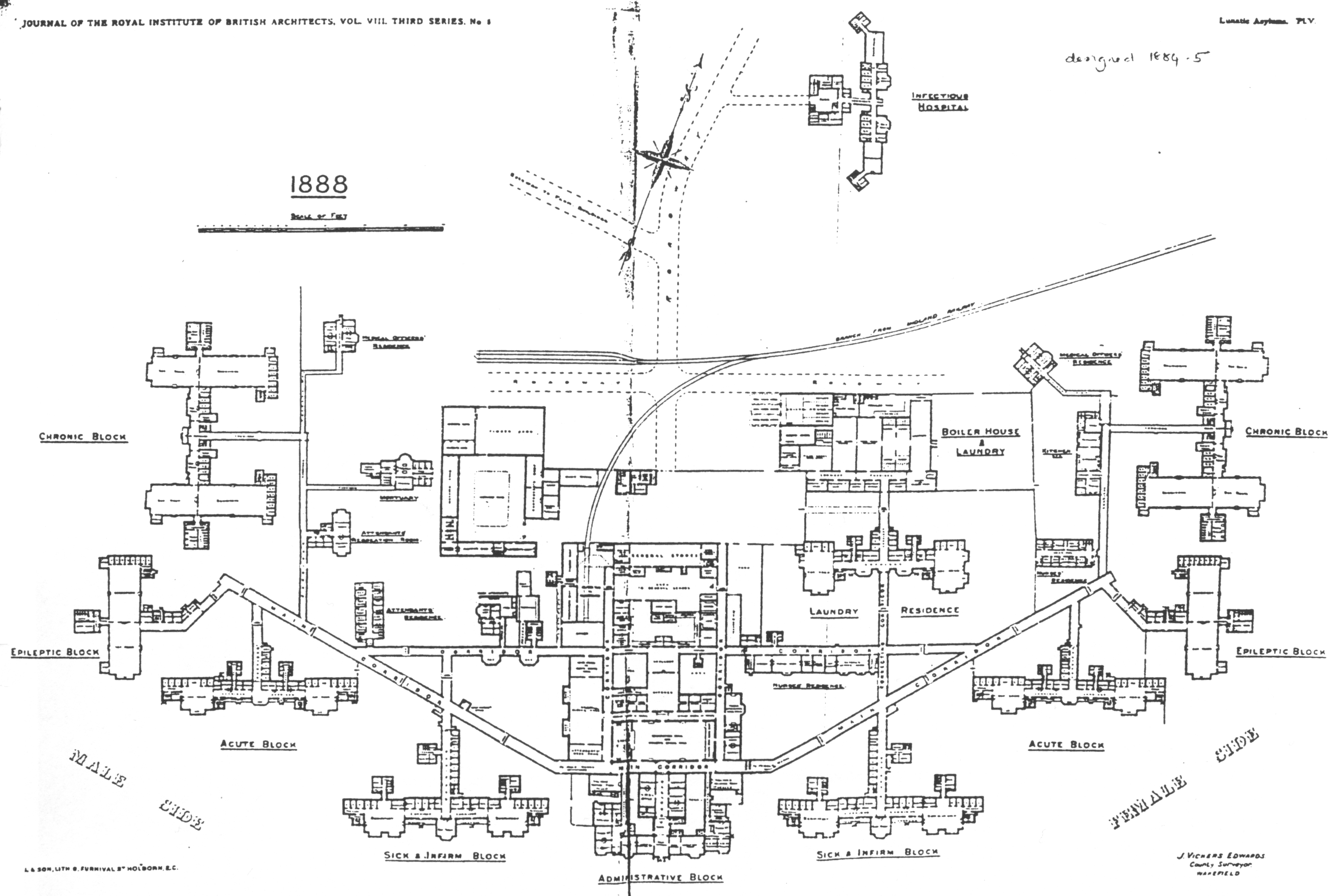
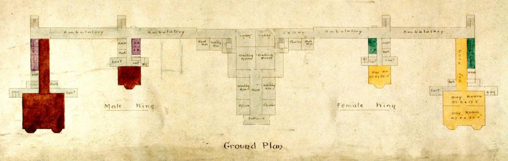
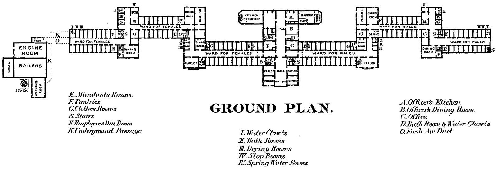
    And so I went looking around the web for other examples of mental/insane asylums, places constructed for the good of the society and partially to aid people who were suffering from some sort of yet-to-be-defined mental illness.  From what I have found so far, there is a definite them to a good many of them, resembling in some fair manner the Morristown design (above). 

    (An interesting and lush/ruinous photo essay on Morristown can be found here.)

    I suppose that in some ways these architectural plans are maps, outlines to an elusive wellness; given the (generally) small staffs, the abilities of the time to diagnosis mental illnesses, the limited capacities for treatment, and general overcrowding, that the “wellness” part of the desired ends of these plans/maps was almost entirely allegorical. I suspect that if for some reason you found yourself in an extended stay in a place like this and weren’t insane, then you would be after a while.

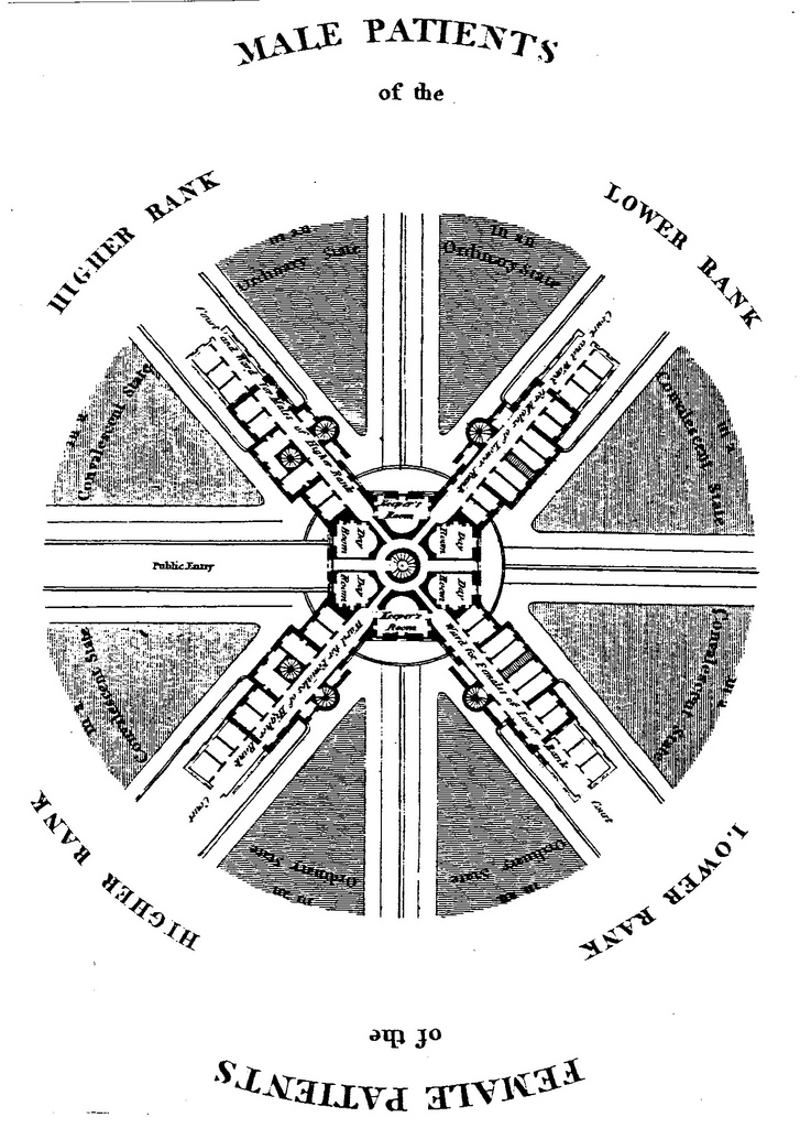
    The Glasgow Lunatic Asylum, designed by the  architect William Stark (1770 – 1814):

    Lunatic  glasgow

    [From:  Thompson, John D. & Grace Goldin. The Hospital. A Social and Architectural History. Yale University Press, New Haven and London. 1975.]


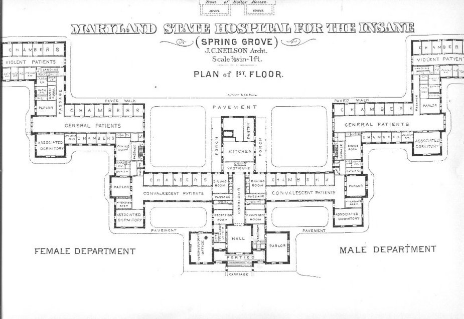
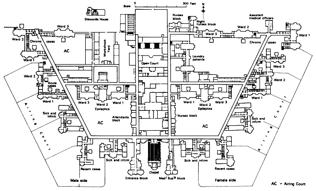
    Insane asylum maryland state hospital

    Maryland State Hospital for the Insane (here)–in this plan, the further away you get from the main entrance, the more deeply “disturbed’ the patients.  It must have been a long walk from the “violent” ward to freedom. 

    Lunatic cane hill

    Cane Hill, the Third Surrey County Pauper Lunatic Asylum, built 1882 (details here)


    Lunatic claybury county

    Claybury Lunatic Asylum, Woodford, Essex:

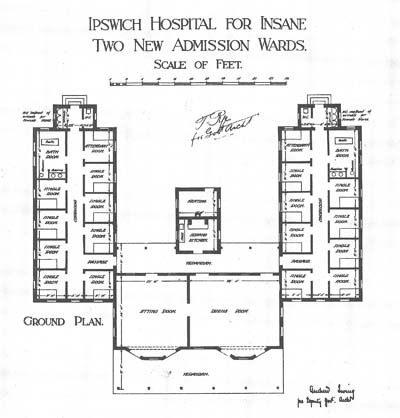
    Insane ipswich

     The Byron House, constructed 1915, at the Ipswich Lunatic Asylum (Byron house was to become the infamous Karrala House.)

    Lunatic buffalo richardson

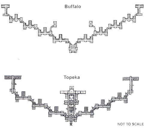
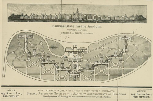
    Buffalo lunatic asylum, a spectacular structure designed by H.H. Richardson, with grounds and landscaping by none other than Frederiick Law Olmstead and Charles Vaux  The image above (and below) are the Kansas State Lunatic Asylum constructed 1880.  Information on these two institutions and the man who brought them about is located at the Kirkbride Buildings blog, here.

    Lunatic kansas


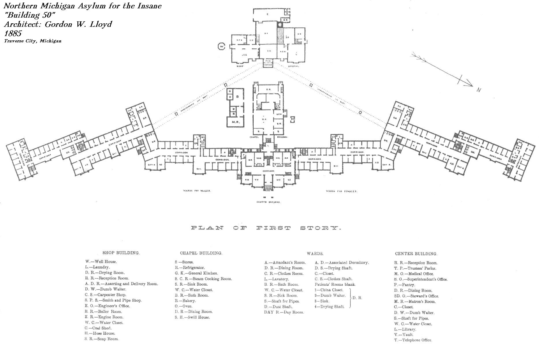
    Northern Michigan Asylum for the Insane, built by Gordon W. Lloyd in 1888:

    Lunatic  northern michigan

    Seacliff Lunatic Asylum, New Zealand:

    Lunatic seacliff


    HArrisburg (PA) State Lunatic Asylum, the first to be built according to the Kirkbride Plan:

    Lunatic  harrisburg

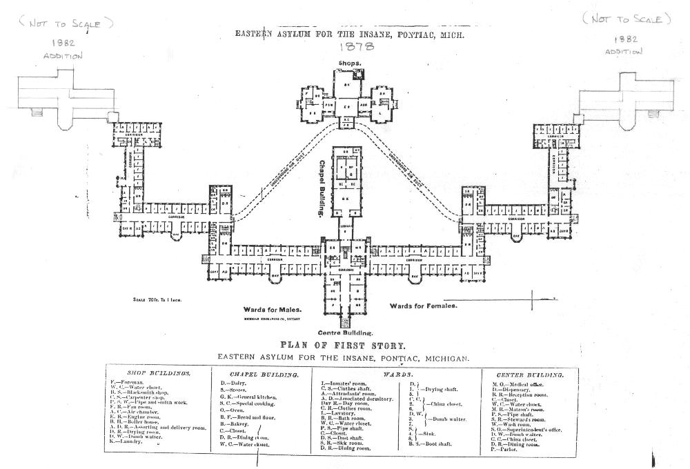
     Eastern Asylum for the Insane, Pontiac, Michigan, constructed 1882:
    Lunatic  eastyern asylum


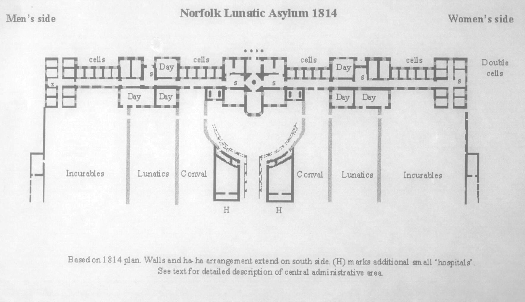
    Norfolk, England, Lunatic Asylum, 1814

    Lunatic norfolk

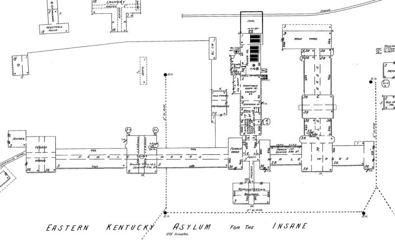
     Eastern Kentucky Asylum for the Insane:

    Lunatic--eastern KY hospital for the insane


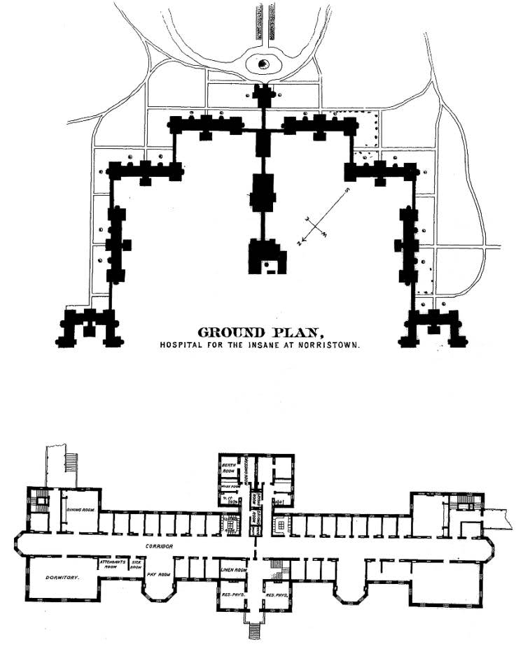
    Norristown Hospital for the Insane:

    Lunatic--norristown hop for the insane