A Daily History of Holes, Dots, Lines, Science, History, Math, Physics, Art, the Unintentional Absurd, Architecture, Maps, Data Visualization, Blank and Missing Things, and so on. |1.6 million words, 7500 images, 4.9 million hits| Press & appearances in The Times, Le Figaro, Mensa, The Economist, The Guardian, Discovery News, Slate, Le Monde, Sci American Blogs, Le Pont, and many other places… 5000+ total posts since 2008.

Category: Medicine, History of

  • Explosive Anatomy, 1833

    JF Ptak Science Books   Post 1479– (from 2011, expanded)
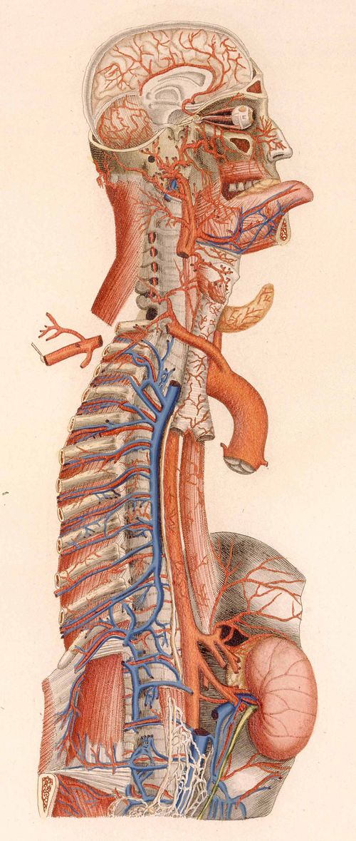
    Anatomy--mascagni[Mascagni image(s) source: National Library of Medicine, here.]
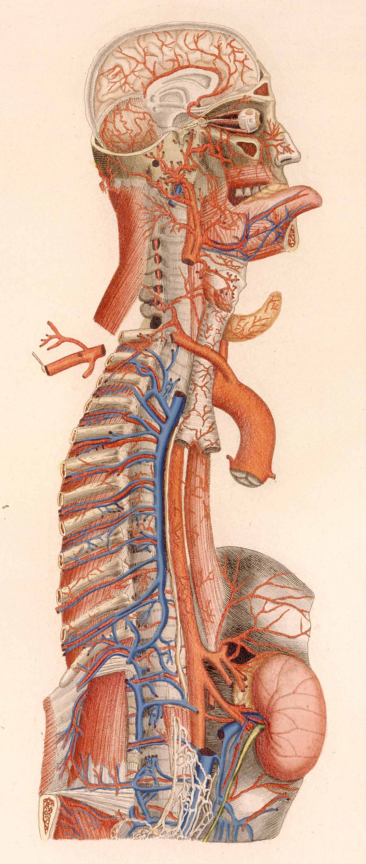
    Species of explosions come in all sorts of shapes and sizes, and basically release some sort of energy (chemical, nuclear, mechanical and so on), but in the cases we’ll look at here they are none-of-the-above.  The energy released in these explosions are in the form of potential, a knowledge-driven, controlled and reversibly ordered demolition, a deconstruction in slow motion.  The medical “explosion view” for the study of anatomy is a remarkable thing, especially when you considered not only the pre-CT and pre-MRI times, but also the pre-X ray (1895) days of imaging the inside of the body–the images not only show you the bits and pieces alone but also in relation to the other bits around them.  The body becomes an archaeological dig, each level preserved so that a viewer could easily see the dependencies and relations between one thing and another. 
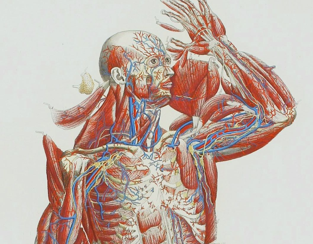
    Anatomy mascagni three

    Take for example Paolo Mascagni’s (1755-1815) beautiful Anatomia universale (an example seen above), which was printed in Florence in 1833 and has the look and sensibility of something much later.  Mascagni is able to achieve this depth not only from the exploded view, but also for the (somewhat exaggerated) coloring setting the specimen on top of a blank background the combination of the color and the background giving the whole thing a 3-D-ish feel–and if not that, then certainly a living image in which there is some fair amount of depth. 
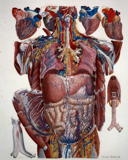
    Anatomy exploded
    Another version of the exploded view which is also quite effective from the same work by Mascagni is his “exploded thorax”, showing a more limited display of the more closely-aligned organs. 

    When these views appear today–which is generally much more uncommon given the other technical ways of envisioning multiple layers of complex associated items–they have more of a sense of the antiquarian, retro feel to them, though they are no less useful for it.  As a matter of fact  I still prefer these views for understanding technical and engineering  and architectural complexes than anything else. And the modern master of this genre for me is Stephen Beisty, who performs his magic mostly in the tech and engineering fields, but who occasionally dips into the messy wet stuff of biology, as we can see in this fabulous exploded view:
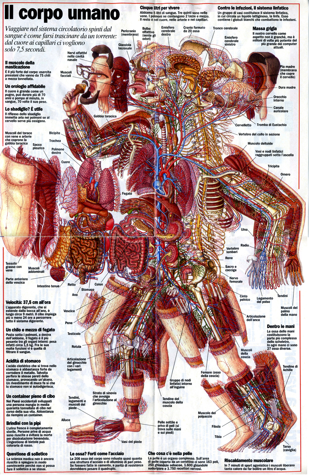
    Anatom exploded beisty

    There are of course many other examples of (drawn) exploded views in the history of anatomical illustration, but I think these will do for now as good examples of the art.


  • Electroshock Therapy for Melancholy in the 40+ Age Group

    JF Ptak Science Books   Quick Post

    Follow Me on Pinterest

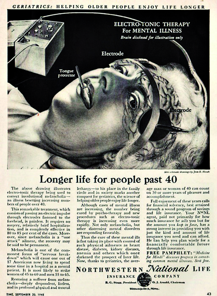
    This advertisement for a remarkable new and ‘painless” therapy for involutionary melancholia looks to me strained and strangely Ken Keasyian, John-Forbes-Nashian. The disease–not actually recognized as such by the DSM-5–was said to be lessened by “electro-tonic therapy”, which looks to me to be electroconvulsive therapy, or ECT.  Evidently before treatment for illness like involutionary melancholia were available via antidepressants the ECT worked in some number of cases.  Still to me this looks like all kinds of bad.  

    ECT therapy

    ·Posted by :

    ·in:

    —

  • Kingdoms of Dust and Street Dirt, and What People Breathed in 1878

    JF Ptak Science Books  Post 2307

    Here’s an extraordinary find, a bump in the grazing field in the Scientific American Supplement for January 19, 1878. The article is about dust–but not the cosmic dust that some folks say is what is being seen in the Big Bang (and not a background radiation signature), nor is it like the dust equation nor the dust of Einstein’s great dust paper of 1905.  It is more like a lower-level plague dust as we see in Arthur Rothstein’s iconic dust bowl photograph made in parched Oklahoma in 1936.  It is about the residue of industry and commerce that did’t get carried of by the mysterious carry-off winds associated with the high and higher chimneys of Victorian London and other industrialized cities, and the leftover bits of the operation of daily cosmopolitan life involving say horses and trains.  It is an article about the significant particulate  matter of dust, which we know today is a high-percentage contributor to air pollution, not to mention the low-level “atmosphere” of the kicked-up business that people would breathe in every day.

    And, evidently, this dust was very heavy-metal rich in composition, which is not so good.  

    The article is “Street Dust”, and the author, Henry G. DeBrunner (who would become professor of chemistry at the Pittsburgh College of Pharmacy), decided to take a look at the dust of Pittsburgh and compare it to reports of the dust of major European cities.  He notes that Paris and London and other cities consist of 35% of “metallic iron, given by the shoes of horses to the stones, besides from 30 to 40% of good glue from the hoofs”.  Now that seems an extraordinary figure, and surprising too in many ways that it came from horse shoes. 

    Dust mask296

     [Source: an advertisment from Scientific American, volume 79, 7 January 1893, back wrapper.

    DeBrunner conducted a study at Thirtieth and Smallman Streets (Pug) and found out that 30% of the sample of dust (“a deep black color”) consisted of silic acid, 26% of “fixed carbon”, ferric oxide at 12%, sulphuric acid at about 1%, gluten 1%, Metallic iron 8.55%.  The details of how all of this was extracted can be seen below, where the entire article is reproduced. There were other samples, and it is interesting to note that in one sample near horse tracks contained 32% heavy metal and lots of glue. 

    The entire article, January 19, 1878, Scientific American Supplement, No. 107:


  • First Printed & Illustrated Medical Book (1500)

    JF Ptak Science Books   Post 2277

    Ketham_p37 det
    “For my part I deem those blessed to whom, by favour of the gods, it has been granted either to do what is worth writing of, or to write what is worth reading; above measure blessed those on whom both gifts have been conferred”–Pliny the Elder

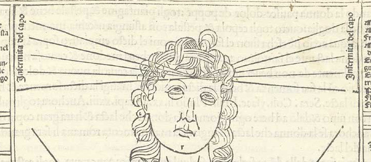
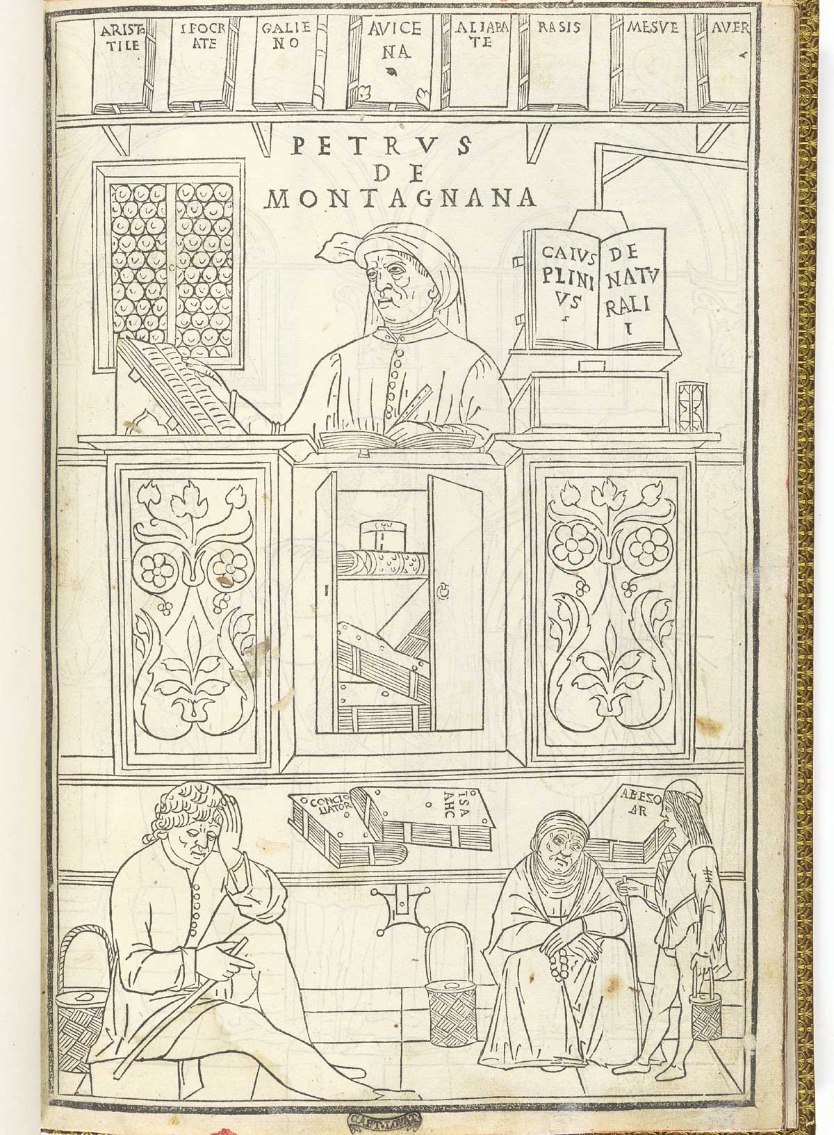
    Johannes de Kehtam’s Fasciculus Medicine (printed in Venice in 1500) was the first anatomy to be printed with illustrations. Ketham was described as a German doctor living in Italy and may well have been Johann von Kerchheim, a German practicing surgery and medicine in Venice during he 1470′), and who wrote a series of tracts on various aspects of medicine which were then collected into this single bound volume.  The illustrations are spectacular and to me have a very modern sensibility in their mid-Renaissance woodcut legacy–the look very clear and concise, are well proportioned, nicely labeled, and give plenty of free rein to open and blank spaces on the woodblock.  The only time these images really “fail” is when they appear in color–a process that would’ve been undertaken privately, by the purchaser of the book, who would have contracted with an artisan to color the book. The images in almost all of the cases of coloring that I have seen just do not match the elegance and brilliance of the original with no color. 

    [Source for all images: NATIONAL LIBRARY OF MEDICINE, here.]

    Ketham_p01

    Its interesting to make a quick note on the books on display on the bookshelf behind the seated figure of Petrus de Montagnan (who is consulting classic texts in medicine as his three patients awaits his attention): the books  include Aristotle, Hippocrates (“ippocrate”), Galieno (Galen), Avicenna (980-1037), Rasis (Ibn Sina, Rasis, Razi, Al-Razi, c.865-925), Mesue, Yuhanna bin Masawayh (c.777-857), and Averroes (1126-98), Abu el-Walid ibn Ruchd, Abu el-Walid ibn Ruchd. On the shelf to the right we see a copy of Naturalis Historia by Cais de Plnii (Gaius Plinius Seocndus, also known as  Pliny the Elder, who lived from 23-79 A.C.E and who held forth on all knowledge from everywhere during the first century).

    It is the anatomical illustration where the un-named artist is most brilliant–the lines are sharp and crisp, the spacing elegant, and the imagery very clear.  It is just lovely work:


  • High Realism in Anatomical Art, 1690 & 1872

    JF Ptak Science Books      Post 2276

     Anatomy--braun--

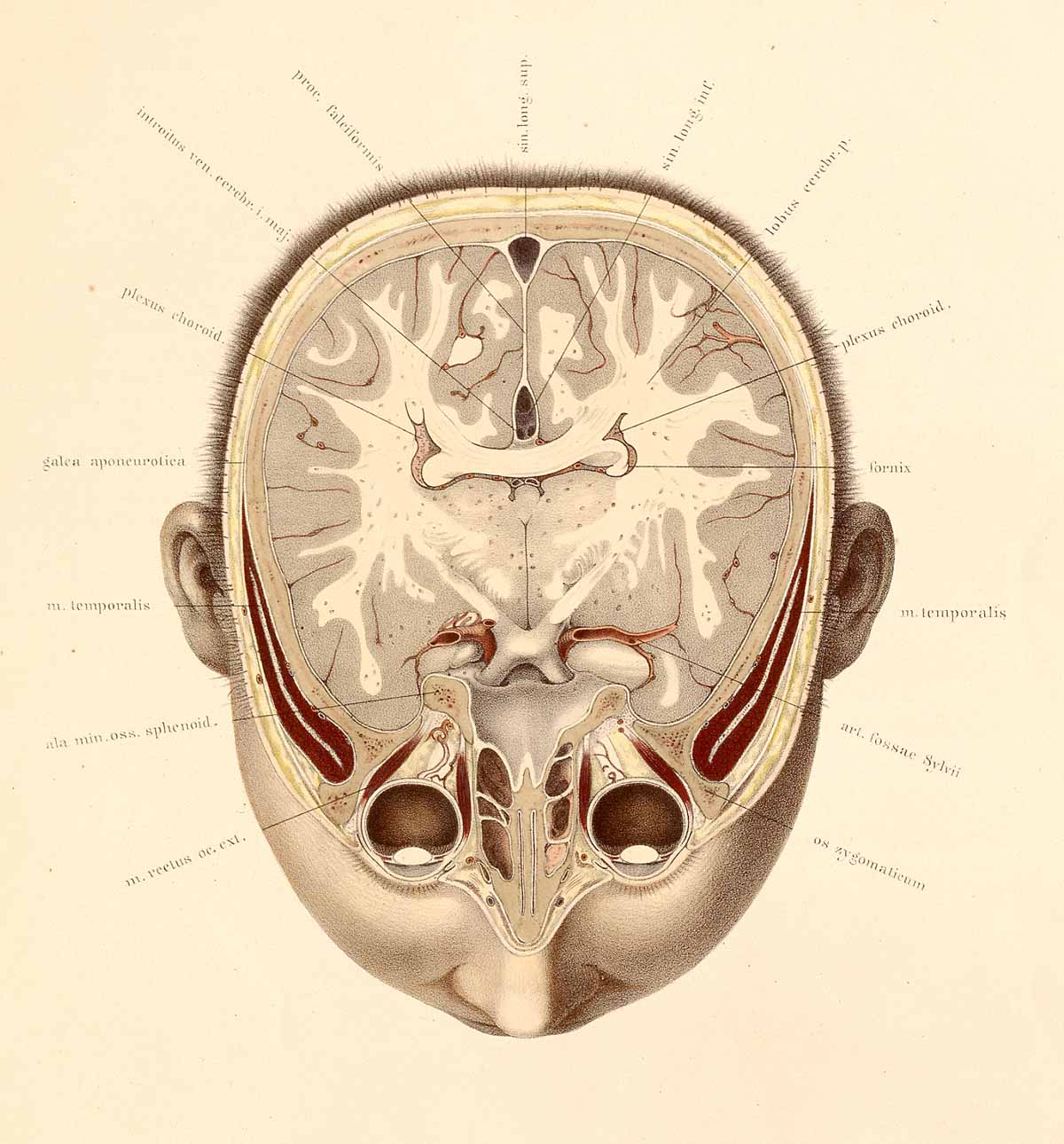
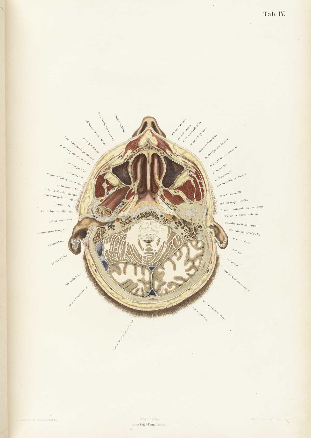
    The collaborative work of anatomist Wilhelm Braune (1831-1892) and artist C. Schmiedel (fl. mid-1800s) produced a singular work,  Topographisch-anatomischer atlas nach durchschnitten an gefrorenen cadavern…(published in Leipzig in 1872), which was an oustanding example of high-realism in the art of anatomy. This book is pretty much right-to-business, with no extraneous bits as had appeared in anatomies fo rcenturies. And even though these practices had pretty much expired by the first quarter of the 19th century, Braune/Schmiedel was ever more so detailed and simple–their design was fabulous, and their detail light and exceptional. 

    Anatomy braune04[Source: National Library of Medicine.]

    They also made use of sliced frozen sections of cadavers, which seems to give their work that crystalline touch.  The first image looks at the brain from top-to-bottom, while the second reverses the view, which is in itself somewhat unusual, even in 1872. 

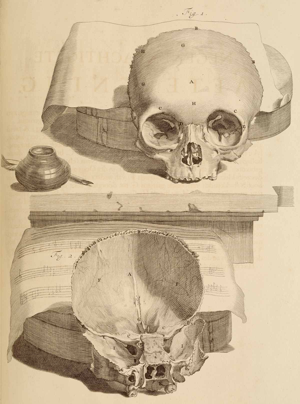
    Looking into a very finely-produced skull from an earlier period in the work of the anatomist Govard Bidloo (1649-1713) and the artist Gérard de Lairesse (1640-1711) in their Ontleding des menschelyken lichaams, (which was printed in Amsterdam in 1690) we find a bit of a different story.

    Anatomy skull[Source: National Library of Medicine.]

    One interesting aspect of Bidloo is that he can be extraordinarily detailed and still lend a rather baroque still life to engage his subject–like the skull with musical score, above.  There’s no reason for the added bit of scenery, its just a luxurious background for the anatomy. And then there are examples like this incredible facial/neck dissection below, which is very hard-core realistic, just as what you might find on any dissecting table, which most assuredly did not come equipped with pen/ink/music score.  Bidloo here is ultra-real, more so than the greater majority of anatomist/illustrators. [Ontleding des menschelyken lichaams...printed in Amsterdam, 1690, as a copperplate engraving with etching. Source: National Library of Medicine.]


  • Affairs of the Heart: Blood & Clouds, 1664

    JF Ptak Science Books  Post 2270

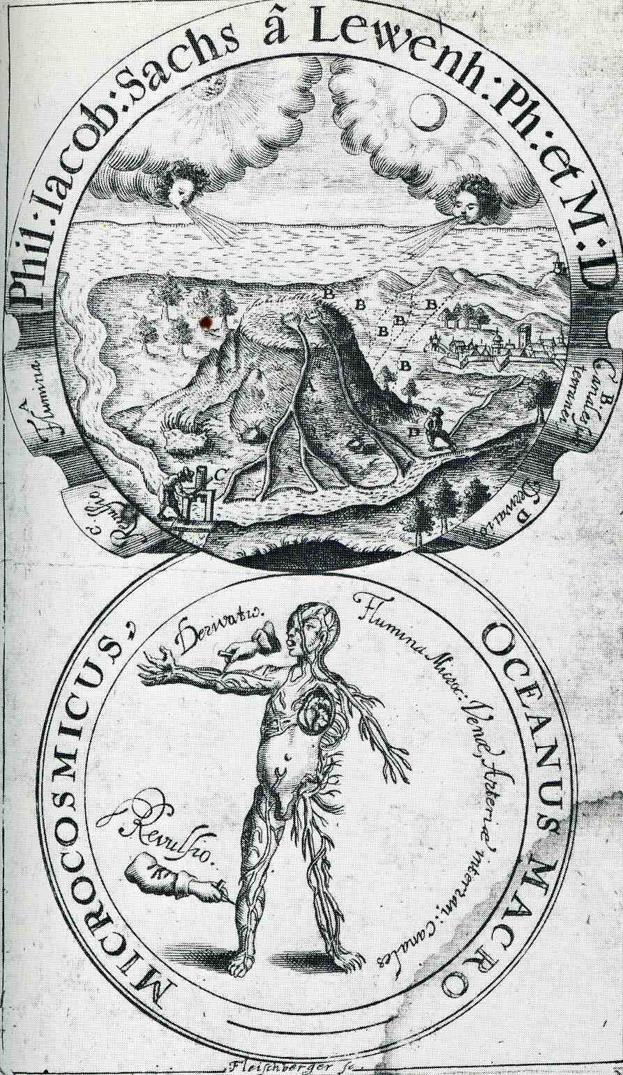
    0 blog feb 6 circle541

    [Image sources: National Library of Medicine.]

    In 1664 Philipp Jakob Sachs (Sache de Lewenbheimb) wrote an influential book on the circulation of the blood.  It was the advanced work of a learned man, a naturalist and physician who was also the editor of the Ephemerides Academiae naturae curiosorum, which was the first journal in the field of natural history and medicine and one of the founders of the Academia Naturae Curiosorum (Leopoldina).  His work came 40 years after the great work by William Harvey, who published Exercitatio Anatomica de Motu Cordis et Sanguinis in Animalibus in 1628, a work in which he essentially brought the idea of circulation of the blood into the modern age, building on earlier ideas of Michael Servetus, whose 1561 work on circulation (and his religious ideas) brought him to be executed by flames. 

    For centuries the pulse was a vaguely understood thing reaching back into the murky medical past as far back as Galen.  The association of course was with the heart, and the association of the heart was as the great controlling center of all function and control of the human body—a theory that reached far forward into the 16th century.

    Servetus (physician, cartographer, theologian, writer and general all-adept Humanist of a high order) was in trouble with the church for many reasons, not the least of which was trying to dislodge the theory of the heart as sacred and the seat of wisdom.  But he did establish that the heart was an organ, which didn’t sit well with very many people, least of all the Calvinist court in Vienna which found him guilty on many anti-Humanist grounds, including his anti-Trinitarian Christology, which made him a reviled figure to Catholics and Protestants.  He was tried and found to be dangerously heretical, and sent to the flames.

    Harvey withstood blistering attacks on his correct statements on the circulation of the blood (costing him nearly all the patients in his practice), though he at least lived to see a brighter day:  Servetus, on the other hand, didn’t, and was burned at the stake for his heresies, one of which his attack on the spiritual heart.

    In any event, the frontispiece to Sachs’ work is an interesting allegorical composition showing a connection between the place of the very prominently featured heart in the circulation of the blood, and the water cycle, and the cosmos of creation (the breath of life coming from the winds of the Sun and the Moon).   

    0 blog feb 6 circle542


    Notes:  (Sachse de Lewenheimb, Philipp Jakob Sachs, 1627-1672 , Oceanus macro-microcosmicus, seu Dissertatio epistolica analogo motu aquarum ex et ad Oceanum, sanguinis ex et ad cor… 1664, with full text via Google books here).


  • Bill of Mortality, London, December 1665

    JF Ptak Science Books   Quick Post

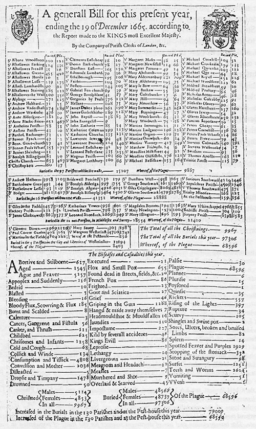
    Six days before Christmas in 1665, the city of London and environs had suffered through more than a year of the Bubonic plague:  the Great Plague of London, killer of 100,000+ people to that point. It was in this preceding week that the annual report of deaths in the city of London were reported in this very grim bill of mortality:

    Bill_of_Mortality

    [Image source:  Wikicommons]

    ·Posted by :

    ·in:


  • A Note on Graphical Display and a Mechanical Heart, 1876

    JF Ptak Science Books  Post 2138
    Follow Me on Pinterest


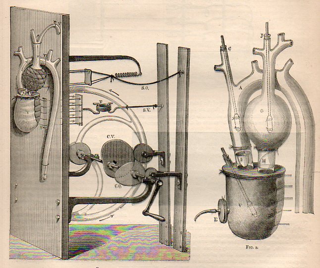
    Marey mechanical heart816

    This quick note comes as the result of a short chase on the detection of the pulse and the ability to determine death–as far back and even before Pliny life was determined by the audible heartbeat, which is where the trouble began, because one can still be very alive with a faint heartbeat. This was a major concern when dealing with folks who were thought to have expired, because in the mid-19th century and before the instruments necessary to make a careful and accurate appraisal of whether the heart was still working were yet finely developed.  Laennec’s stethoscope appeared around 1816, but as much of an improvement as it was this instrument awaited considerable refinements before a truly solid identification of a non-working heart could be established. The gaps in the determination of the arrival of death led to mortuaries where the supposed-dead were left to themselves at room temperature for three days, awaiting the ultimate determination of death which was putrefaction (as in the Munich and Frankfurt Leichenhaus and the Vienna  Zentralfriedhof). Earlier in the history of the determination of death methods were quicker and more brutal (if the patient was still alive) involving bellows-driven tobacco-smoke enema,  as well as tongue pulling and nipple twisting.  (See an earlier post here on the Worst Jobs of the Nineteenth Century for more on this.)

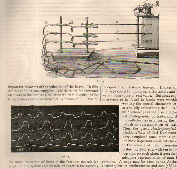
    Marey mechanical heart817

    This led to Etienne Marey (1830-1904), a versatile experimenter and premier instrumentalist who was a scientist, physiologist, and motion-picture/chronographer pioneer, who in the 1880’s created what was essentially the world’s first moving-photographic “slow motion” device. One iteration of Marey’s apparatus was basically a long series of ganged cameras recording a motion for a  simple task at a given time frame and presented on a continuous strip of photographic paper, sort of like a motion picture with the camera speed set at three frames per second. The resulting images were phenomenal and showed people for the first time the exactness of all manners of simple motions–motions that no longer looked so “simple” once all of its aspects could be studied from captured photographic evidence.  Even the act of hopping over a small stool or bending to pick up a bucket of water were enormously revealing in a way like Robert Hooke’s Micrographia displayed the great detail and complexity of the seemingly simple fly.

    Aside fron being one of the founders of cinematography, the other aspect of Marey’s interest in capturing and manipulating time was in medicine, where by the time this article of interest in this post was published in 1876 he had already established himself as one of the greatest cardiovascular physiologists.  What we find in this review in Nature (Thursday, January 6, 1876) of Marey’s Physiologie Experimentale (Paris, 1876) was the editor’s keen interest in the mechanical heart Marey had constructed to show the actions and functions of the heart–the first time, the article notes, that all aspects of the action of the heart were exhibited correctly in one model. 

      Of particular interest was the recording device for the pulse of the heart, which was one in a series of devices such as that improved upon, something Marey himself had done with the first-introduced sphygmograph (“pulse wrtier”) of Karl von Vierdodt in 1854.  The 1875/6 Marey instrument made major advancements in continuous graphical registration in instruments of continuous noninvasive arterial responses.


  • Uncomfortable Dissection Placements of Dissected Material(s), 1546

    JF Ptak Science Books   Post 2131 
    Follow Me on Pinterest

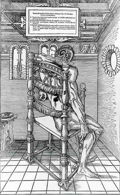
    0 blog dec 27 estienne dissection323

    These remarkable woodcut images come from Charles Estienne’s (1501-1564) De Dissectione Partium corporis Humani linri tres,
    a 375-page opus illustrated with 62 fantastic full-page woodcuts and
    published in 1546  This work is surpassed I think only by the
    masterpiece Fabrica by Vesalius (1543)–the great standard of anatomical illustration.

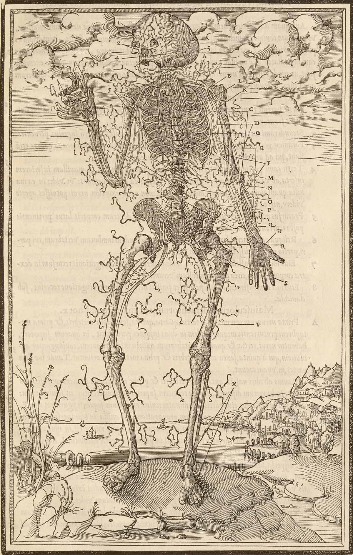
    The images are remarkable not only for their superior and significant anatomy, but–for me–also for the placement of dissections with the body from which they were taken. 

    The particulars:

    Artist: La Rivière, Étienne de, d. 1569. Engraver: Jollat, Jean “Mercure,” fl. 1530-1545.

    Title: De dissectione partium corporis humani libri tres.

    Publication: Parisiis: Apud Simonem Colinaeum, 1545.

    The entire series of illustrations from the National Library of Medicine, here.

    Estienne

    ·Posted by :

    ·in:


  • Affairs of the Heart: Blood & Clouds, 1664

    JF Ptak Science Books  Post 2025 

    Follow Me on Pinterest 

    0 blog feb 6 circle541

    In 1664 Philipp Jakob Sachs (Sache de Lewenbheimb) wrote an influential book on the circulation of the blood.  It was the advanced work of a learned man, a naturalist and physician who was also the editor of the Ephemerides Academiae naturae curiosorum, which was the first journal in the field of natural history and medicine and one of the founders of the Academia Naturae Curiosorum (Leopoldina).  His work came 40 years after the great work by William Harvey, who published Exercitatio Anatomica de Motu Cordis et Sanguinis in Animalibus in 1628, a work in which he essentially brought the idea of circulation of the blood into the modern age, building on earlier ideas of Michael Servetus, whose 1561 work on circulation (and his religious ideas) brought him to be executed by flames. 

    For centuries the pulse was
    a vaguely understood thing reaching back into the murky medical past as far
    back as Galen.  The association of course
    was with the heart, and the association of the heart was as the great
    controlling center of all function and control of the human body—a theory that reached
    far forward into the 16th century.

    Servetus (physician, cartographer, theologian, writer and
    general all-adept Humanist of a high order) was in trouble with the church for
    many reasons, not the least of which was trying to dislodge the theory of the
    heart as sacred and the seat of wisdom. 
    But he did establish that the heart was an organ, which didn’t sit well
    with very many people, least of all the Calvinist court in Vienna
    which found him guilty on many anti-Humanist grounds, including his
    anti-Trinitarian Christology, which made him a reviled figure to Catholics and
    Protestants.  He was tried and found to
    be dangerously heretical, and sent to the flames.

    Harvey
    withstood blistering attacks on his correct statements on the circulation of
    the blood (costing him nearly all the patients in his practice), though he at least
    lived to see a brighter day:  Servetus, on the other hand, didn’t, and was
    burned at the stake for his heresies, one of which his attack on the spiritual
    heart.

    In any event, the frontispiece to Sachs’ work is an interesting allegorical composition showing a connection between the place of the very prominently featured heart in the circulation of the blood, and the water cycle, and the cosmos of creation (the breath of life coming from the winds of the Sun and the Moon).   

     0 blog feb 6 circle542

    Notes:  (Sachse de Lewenheimb, Philipp Jakob Sachs, 1627-1672 , Oceanus macro-microcosmicus, seu Dissertatio epistolica analogo motu aquarum ex et ad Oceanum, sanguinis ex et ad cor… 1664, with full text via Google books here).

    ·Posted by :

    ·in: