A Daily History of Holes, Dots, Lines, Science, History, Math, Physics, Art, the Unintentional Absurd, Architecture, Maps, Data Visualization, Blank and Missing Things, and so on. |1.6 million words, 7500 images, 4.9 million hits| Press & appearances in The Times, Le Figaro, Mensa, The Economist, The Guardian, Discovery News, Slate, Le Monde, Sci American Blogs, Le Pont, and many other places… 5000+ total posts since 2008.

Category: Medicine, History of

  • Bad Resurrections: 19th Century Cadaver Trade in New England

    JF Ptak Science Books LLC  Post 585       Blog Bookstore
    History of Missing Things Part XXXVII

    Grave rob For some, “anatomy is
    destiny”, but not in the way the reach-y Sigmund Freud meant–their
    anatomy quite literally was their destiny, their bodies resurrected (in the bad
    way) from their coffins by chin hooks and sent to the local med school for
    quick and dirty study.  This night-time
    version of ice fishing for cadavers was addressed in a sniffily conceited but
    informative book by A.M. Lassek, Human Dissection:  Its Drama and Struggle (1958).  Lassek, professor of anatomy at Boston University ,
    presides over all aspects of the history of anatomy, but I somehow found my way
    instantly to the cadaver-catching mechanical operations end of it: specifically
    the work of American specimen providers in
    in the early part of the 19th century. 

    Listen:  if I found my way back in time in the Boston area in 1835 and
    was the night jobber and bad-‘un for the local apothecaries hunting down the
    lonesome freshly dead, I think–with praise to Lassek–that I’d be
    able/equipped to do the job.  I hadn’t
    really thought very deeply about the supply of cadervous specimen for the
    thriving medical schools before, but there are many considerations to the
    proper way of stealing a body. (And by “proper” I mean “not
    being caught doing it”.) First of all, there was a serious spike in the
    creation of medical schools, the number of recognized institutions in America
    increasing from seven in 1810 to 33 in 1840 (and 90 in 1870), with a huge new
    population of data-thirsty students to go along with it.  Human anatomy was an exceptionally difficult
    subject to demonstrate with actual specimens (due to moral and legal
    restraints), so the med schools (including the Harvards) had to resort to
    creative solutions:  the bodies had to be
    “found”. And finding bodies had more to do with stealing them than
    arranging for the corpse of a condemned criminal to be purchased from the
    sheriff. 

    The local townsfolk and neighbors of
    medical schools knew this all too well. 
    There were a number of precautions that the bereaved could undertake to
    prevent the resurrection of their departeds. 
    One simple way was to have a guard at the grave all night long–that
    would certainly cause some pain and consideration for the grave robber, though
    it has been the case where the guards were plied with money and alcohol to
    effect a soft gaze on the grave.  Since
    the newly interred laid under six feet or so of newly churned earth, there was some
    amount of extra stuff that was thrown into the pit.  Stones, and rocks and tree limbs were added
    to the hole to make a tougher and noisier go for the robbers; also, wide heavy
    planks were placed width-wise across the coffin and then covered so that the
    robbers would have to dig much wider in order to pull up the boards before
    getting to the coffin.  Another fantastic
    bit that I had never thought of but which falls into the category of branding
    cut lumber for the ride down the river: 
    intricate designs were placed on top of the grave with sticks and stone
    and such so that the family members would be able to detect a defiled
    grave site.  All of this attention would be
    heaped on the grave site for ten days or so–after that the body was deemed too
    far deteriorated as to he of any use to the medical school.

    “Detect?”  Yes, indeed. The grave robber needed to be
    stealthy, leaving no indication of the dark and stinking deed.  Working in very low or no light, the robbers
    would find their mark and lay at least two tarpaulins on the ground. they would
    collect all of the dirt in these catch-alls so as to not excite any attention
    that the grave had been disturbed.  They
    would dig a 3×4 foot hole towards the head of the casket, and, once reaching
    their prize, would drill holes (quietly!) into the wood, break it, and then
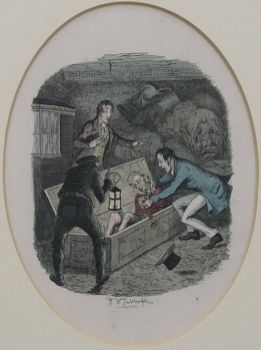
    attach a hook under the chin of the corpse and yank it out.  Placed on another tarp, the body would be
    stripped of all clothing, and then wrapped securely to prevent anything
    incriminating falling from it during the trip across town.  Once received by the medical school, the
    hair, teeth and other distinguishing characteristics would be removed to
    prevent detection and identification.  I
    imagine that the skin of the face would also have been peeled away, skillfully
    removed with one cut and a careful pull. 
    All of this was precautionary, in case the relatives of the now-specimen
    suspected foul play and came looking for their beloved.  It would be a corpse among other
    corpses.  Or not. Lassek writes that the
    bodies would be hidden, secreted in different and obscure places around the
    campus, in “domes, belfries and observatories”.  Observatories?  Well! 
    I’m not so sure about that aspect of Lassek’s report on grave robbing,
    but I was pretty impressed with his knowledge on the other aspects of the
    business. 

    I am
    indebted to Dr. Lassek for the image of the grave-marking anti-theft
    artwork.  I may have fabricate a grubby
    1837 cemetery keeper’s chapbook with drawings of their designs and make an
    entry for it in my imaginary museum catalog for
    things-that-don’t-exist-but-should.


  • (Un-?) Necessary Emotion: Medical and Anatomical Illustration and the Human Spirit

    JF Ptak Science Books LLC  Post 565

    Standard illustrations that give a little more than they need to have always been very interesting to me–sometimes this takes the form of completely unnecessary detail in almost-invisible expendable space fillers (as I’ve looked at in this series)

    Gersdorff_-_Schädelwunde

    and sometimes it is in the most obvious places. 

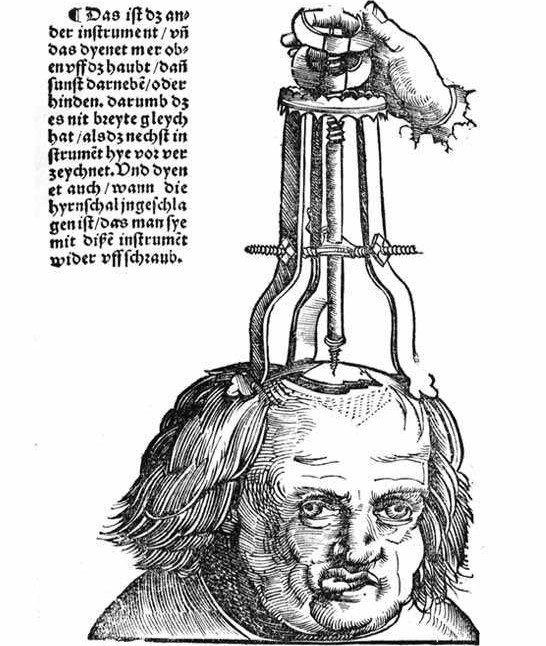
    Now, in this remarkable image from Hans von Gersdorff ‘s (who we met earlier in this blog) Feldbuch der Wunderartzney (Fieldbook of Wound Surgery, and printed in 1517) comes an unexpected bit of detail.  The machine, the surgical device, operates something like a cork-puller: it is meant to attach to and extract a piece of skull that had become embedded in the brain following a skull fracture.  The procedure looks as though it is just beginning–the man’s head has been shaved, the fracture revealed, and the penetration about to begin.  The patient looks, well, I’m not so sure exactly what this look means–perhaps nothing, perhaps the man has brain damage.His right eye has drifted, his jaw set off to the the side and is jutting considerably.  I suspect that a simple view of the top of the patient’s head would’ve been enough to illustrate the image, but–as seems to be more often than not in the 16th century–the illustrator has included not only the entire head, but has given the head character, history, emotion and dignity.  Everything necessary to imbue the surgeon-reader with with the sense of humanity associated with his trade and task. 


    Zzzzzzbartisch

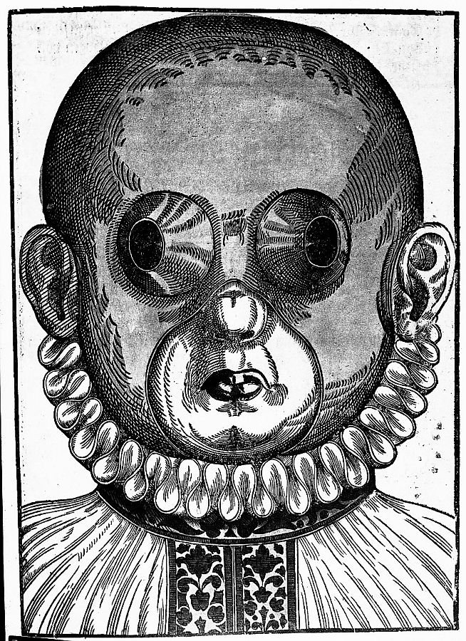
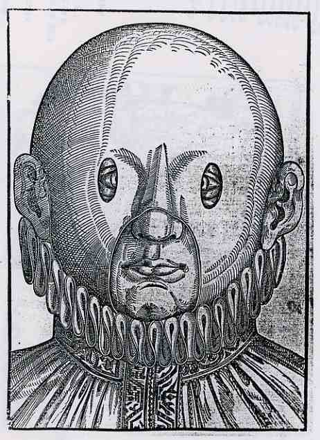
    I should think that the same may be said for many of the illustrations from Georg Bartisch’s Augendienst (The Treatment of the Eye, printed in 1583).  The woodcut at right is an example of Bartisch’s suggestion for crossed eyes–the mask would be worn for different periods of time, the openings supposedly working to make the eye muscles assume their normal positions.  The remedy looks bizarre (and very symmetrical) from our point of view here in its future, but what seems very modern is the careful treatment of the illustration of the patient, who is erect, calm, well dressed and terribly decent. 

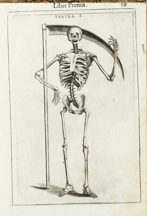
    The thread of deeply humanized anatomy runs deep in the 16th and 17th centuries,where human bodies are dissected to show the different levels of our delicate existence.  The bodies are not simple cadavers, but seem to have some sort of “liveliness” in their post-living being.  This image (below) by Guido Guidi (1508-1569).in his De anatome corporis humani libri
    VII.

     (printed in Venice in 1611)  gives the beckoning skeleton a smile  to go along with its timeless  gesture for the remembrance  of mortality.

    Blog--emotion--guidi 

    Certainly there can be no escaping the modesty of the model
    in Alexander Read’s (1580-1641). Somatographia Anthropine, or a Description
    of the Body of Man. . .By Artificiall Figures representing the members…
    (printed
    in London by Jaggard
    in 1616).  Even though she is displaying most of her abdomen, this person is still caring for her moral appearance.

    Blog--emotion--woman

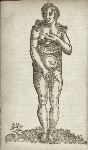
    Another example of this modesty can be found in the work of Adriaan
    van de Spiegel’s(1578-1625). Opera quae extant omnia (edited by Johannes
    vander Linder) printed by the famous Blaeu’s in Amsterdam
    in 1645:  here we see the nearly full-term foetus ably
    displayed, the mother’s skin pulled back as if to form petals to the flower of
    the child (helped along by a strategically-placed plant in the foreground).


    These images can go on and on–these are just a few scant examples.

    Another quick example: 
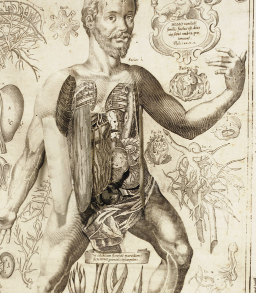
    Johannes Remmelin’s (1583-1632) Kleiner Welt Spiegel, das ist,
    Abbildung goettlicher Schoepffung an des Menschen Leib….
    (printed in Ulm
    in 1661) has many illustrations in which the anatomical model seems quite
    serene; ambiguous, sometimes, to what is going on, but at the same time looking
    inviting, welcoming even.

    Blog--emotion man beard 
    All I’m saying is t hat there seems to be something special going on in these images–certainly a “something” that was lacking in most other (non-artistic) anatomies of the 19th and 20th centuries.  Emotion was certainly extracted from the examples of modern and recent-past anatomical studies. I feel a little happier with the artists and physicians who were interested in connecting humanity to these studies than the slabs of Netter and Grey, to say nothing of the Adam and Eve project or deeply modern technologies.  A little reminder now and then that there was something else connected to the viscera and bones and muscles and veins might not be too bad. 


  • What Pictures Tell Us: Medieval Treatment of Women in a Renaissance World

    JF Ptak Science Books LLC  Post 553

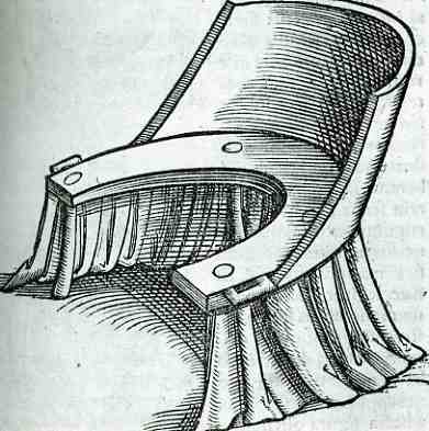
     blog 19 obst chair711
    There are all sorts of famous seats:  seats of wisdom, of power, of lameness, of government, of discontent, of honor, of learning, of war; there’s the hot seat, the best and worst seats, a seat on the board, a seat on the bench, a seat at the bar; there are captain’s chairs, endowed chairs, and famous chairs (like architectural gems/mules of Mackintosh, Wright, Saarinen, van der Rohe, Eames, Gehry, LeCorbu) and famous seats (at the original Yankee stadium or a Roman coliseum).  The seat pictured at left, though, is anonymous and lonely, but it is full of a story. 

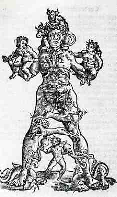
    Sometimes one discrete, odd image can tell you a lot of history or the story of its time:  It might seem simple and benign, but once you stop to think about it just  little, an entirely different story can emerge.  Take for example this woodcut from Jacob Rueff’s De conceptu et generatione hominis, et iis quae circa haeac potyissimum consuderantur (in six books), published in Zurich by Christoph Froschauer in 1554.  It looks like it might be a seat straight out of an old WC, but it is actually a birthing  blog 19 obst astrolog712chair.  And it doesn’t look particularly inviting (or not threatening) for the tasks to come; and such was the state-
    of-the-art of birthing children in 1554.  Well, earlier than that, really, because Rueff’s work was an improved version of a still earlier work by Rosslin (Der swangern frauen). I look at this chair/seat, and I think,  “oh dear god”, even with an historian’s sensibilities, knowing full well that it hadn’t been changed much for hundreds of years prior to this publication.  There’s quite a bit in this book’s 66 illustration to excite the historian of medicine, some of which are rather beautiful and demanding, like this astrological representation of anatomy (at right).  But it is still the chair in all of these pictures that is the most powerful to me. When all of the medical advancements and distractions were boiled away, and the discussion of the two types of foreceps was eliminated, it all came down to a woman in an uncomfortable, stiff-backed straight-up chair.   A chair with no cushion or throw or anything of any sort for the comfort of the woman, though there was an extensive dust ruffle flanking it.  And as great as the advancements were in medicine in the first part of the  16th century, dealing with women in labor was still a Medieval practice.

    I should also point out that the Rueff book, as old as it was, was reprinted, translated, into English with many of its ideas riding out time intact, published in London eight decades later.  The chair was still there. 


  • A History of Missing Dots: Donne’s (Missing) Discovery of Platelets, 1842

    JF Ptak Science Books LLC  Post 547Blog 13 donne detail

     blog 13 donne703 

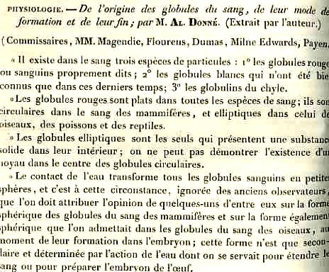
    Alfred Donne (1801-1878), physician, experimenter, microscopist and photographer, is best known for things other than what he should probably be best known for:  his discovery of the third element of the blood, platelets.  Donne had a wonderful vision, and was among the very first on the scene to write about and employ the spectacular invention of the daguerreotype in chemistry and microscopy.  As a matter of fact Donne, along with his collaborator Leon Foucault,  produced the very first engraved images of photomicrographs for his cytology paper of 1840. (He was also the first to use electricity in producing a medical illustration, and was among the first modern physicians to write on the great efficacy of mothers using their own milk in breast feeding their children.)  The discovery of platelets paper of 1842*–preceding two other works published in the same year–does indeed identify the new object, but actually fails to make a methodological examination of the new body, calling the new units “globulins du chyle” (or small globules
    derived from plasma).  He also fails to make a drawing of what he saw; so, the combination of these two important elements asked the reader to accept his findings on faith, the tools of reproduction of his observations not being present.  And so we remember Donne for some other significant things, na dless so for his lightly-interpreted and un-illustrated “dots”.

    *Donne, Alfred. De l’origine des globules du sang, de leur mode de formation et de leur fin. Comptes rendus de l’Académie des Sciences, Paris, 1842, 14: 366-168. 

    (Overall the history of the
    discovery of platelets is convoluted: 
    Leewenhoek (1675) and Henson (1782)  were the first to fully describe the “undefined”
    particles of the blood; Donne’s work was then more fully described by Beale in
    1850, and then again (identified as “small corpuscles” by Zimmerman in 1860 and
    then again by Schultze in 1874 and Laptschinski (also in 1874). William Osler
    then enters the scene in 1880, followed by Giulio Bizzozzero who was the
    first, in the years 1881 -1882, to establish central role of platelets
    not only in physiological haemostasis, but also in thrombosis.  Anyway it is a longish and not-clear
    history.)

    .


  • Lethal Teeth & Causes of Death in London; Milk Leg, Breakbone Fever and Eel Thing. The Death List for 1665

    JF Ptak Science Books LLC  Post 464

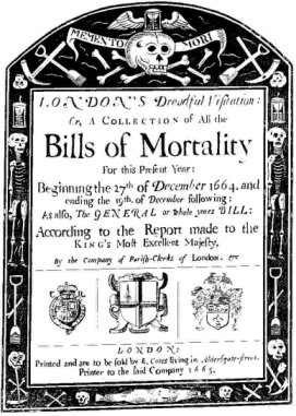
    Blog--bills of mortality
     Coming on the heels of the only post that I’ve made on teeth is this happy
    element, a brief mention of lethal teeth in London in 1665. The listing of the death and
    its introduction (if not the actual dead themselves) comes in the form of a
    publication called The London Bills of Mortality, which was begun (so it seems)
    in 1538.  As an instrument of society the
    Bills were more for legal ramifications than for compiling medical
    information.  The Bill was a necessary
    element for legal burial and the transfer of an estate, composed to make sure
    (or for extent possible at this time) that death had indeed occurred and that
    the legal course could be enacted to disassemble the accumulated physical life
    of the decedent.  The weekly reporting
    and compilations must’ve been wearying, and I imagine that this was not the
    most auspicious or desirable job to have, wringing in the newly dead.  Death’s accounting procedure grew more
    systematized as time wore on, getting a more formal approach as early as
    1603.  And it was not in every English parish
    that the cause of death was recorded, and the recorders were hardly a army of medical
    experts—causes of death were sometimes accurate, sometimes not, sometimes
    whimsical and sometimes all or none of the above.

    The list below attests to a somewhat poetical bent in the naming of lethal
    afflictions; of course most meant something immediate and understandle to the
    reader in 1665, though today many of the names seem quire airy and adventurous,
    hardly things to actual die of.  The list
    comes from the groundbreaking work of London haberdasher and man of
    semi-science, John Graunt, who in 1662 published Natural and Political
    Observations … on the Bills of Mortality
    (1662):  it was a tremendously important work and a
    foundation stone in statistical applications in health and epidemiology, as
    Graunt tried to organize and interpret the collected data. 

    Again, my attraction to this all stems from yesterday’s post on the question
    of the existence of teeth in the Renaissance—“teeth” is listed as a cause of
    death in 39 of these cases, the 6th leading cause of death among the
    39 causes listed.  I guess death might’ve
    been caused by a systemic infection caused by a rotten tooth ; or  perhaps the tabulator observed a dead body,
    saw a mouthful of bad teeth and nothing else obviously wrong with the body, and
    thus dubbed the cause of death as the most heinous looking thing that could be found.


    Abortive
    Aged
    Ague
    Apoplexy
    Asthma
    Bloody Flux
    Cancer
    Childbed
    Chrisoms
    [1]
    Colick
    Consumption
    Convulsion
    Dropsie
    Evil
    Fever
    French-Pox
    Gangrene
    Gout
    Griping in the Guts
    Headmouldshot
    [2]

    3
    52
    12
    1
    2
    1
    4
    4
    2
    2
    78
    112
    26
    1
    76
    2
    1
    1
    14
    2

    Jaundies
    Imposthume
    [3]
    Mortification
    Plurisie
    Rash
    Rheumatism
    Rickets
    Rising of the Lights
    [4]
    Rupture
    Small-Pox
    Stilborn
    Stone
    Stoppage in the Stomach
    Suddenly
    Teeth
    Thrush
    Tissick
    [5]
    Twisting of the Guts
    Ulcers

    1
    2
    7
    2
    1
    1
    1
    1
    1
    54
    9
    2
    2
    1
    39
    1
    5
    6
    1

    A few of the obscure terms explained:

    Ague: 
    deep fever
    Body Flux: sepsis? 
    Childbed: 
    a baby or mother who dies during childbirth
    Crisoms: 
    refers to a white baptisimal robe worn by an infant, meaning that this
    was a very young baby born    before baptism occurred.
    Dropsie: 
    lethal inflammation or edema
    French-pox: 
    syphilis (called “French” because of a decimating outbreak of teh
    disease in the French army.  That, or the
    prescribed treatment, which was the ingestion of mercury. 
    Headmouldshot: edema of the brain
    Imposthume: Abcesses or cysts
    Rising of the Lights:  inflammation of the lungs or general lung
    problesms
    Thrush—an infection of the mouth (especially
    in babies).
    Tissixk: 
    tuberculosis

    According to the website Magic-Nation “other causes of death included affrighted, bladder in the throat, breakbone
    fever, canine madness, commotion, eel thing, frogg, gathering, grocer’s itch,
    hectic fever, kink, milk leg, screws, stranguary, stuffing, rag picker’s
    disease, St. Anthony’s fire, tympany, worm fit, wolf, and being planet struck”.
    Most of these I have little  insight on,
    and even though they caused someone’s death, they still have a lilting, lyrical
    quality about them.  I particularly like
    and am curious about “eel thing”, “milk leg”, and “Stuffing”


  • A Unique Position for Brain Dissection: Estienne’s near-immortal classic (1545)

    JF Ptak Science Books LLC  Post 441

    0 blog dec 27 estienne dissection323
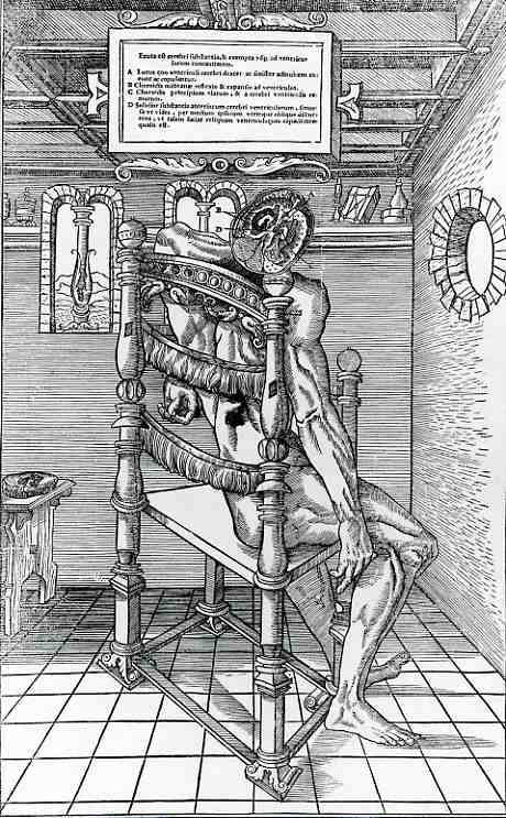
    This remarkable woodcut image comes from Charles Estienne’s (1501-1564) De Dissectione Partium corporis Humani linri tres, a 375-page opus illustrated with 62 fantastic full-page woodcuts and published in 1545  This work is surpassed I think only by the masterpiece Fabrica by Vesalius (1543)–the great standard of anatomical illustration.  Actually, historians have determined that the Estienne book was about ready to go in 1539, but due to a variety of problems, was not published until two years following his fellow-classmate’s Vesalius’ work.  Had things gone a little differently for Estienne, he would most certainly enjoy more ethereal fame than he does now, though I’m sure some of that would be diminished by the weird and exotic nature of many of his prints.  Vesalius chose a somewhat more pragmatic approach, though just about everyone would seem so compared to Estienne.  The Estienne work comes at the beginning of the wave of spectacularly illustrated early books:  Gesner’s 1553 Historiae animalium, Georg Agricola’s De re meteallica (1556), Pierre Belon’s two works on fishes (1555) and birds (1556), Jacques Dalechamps (1513-1588). Chirurgie Françoise… avec plusiers figures des instrumens necessaries pour l’operation manuelle.(1569).and  Matteo Realdo Columbo’s (ca 1510-1559). De re anatomica libri XV (1559). are notable examples of defining-efforts of this period, classics of thought to this day.  And beautiful.

    The beautiful mind here in Estienne’s effort is perhaps the most lovely of all of his illustrations–it is also the most unusual, and the most perspective-abusing.  Our subject is seated in a seemingly-normal armchair of the period, except that the posts lack arms.  The perspective is such that the chair back seems to alternate position, an apparent illusion, changing from front-to-back and back-to-front, while the chair back posts seem to also alternate heights.  Nothing else seems to quite work either:  the floor is at odds with the walls and ceiling, the window is falling inwards, and the chair (again) seems to float when you look at the ceiling.

    Be that as it may, the dissection of the brain takes place with the cadaver in the chair, the body held there with pins (one of which is seen coming through the right shoulder.  The four elements of the brain identified A to D are explained in the ornamental legend at top.  We can see the rest of the top of the head placed on the stool at left, just in case anyone was wondering.

    Again, the Estienne book was a magnificent and somewhat twisty-unusual accomplishment.  Had he published in 1539, which was a definite “maybe”, it would be Estienne who would’ve been known for introducing such things as the first time the entire human body was discussed and illustrated, and the first time that a serial dissection was presented and illustrated.  But he didn’t get the job done in ’39, and so he is not known for these things.  But almost.

    Something happened to Estienne in 1561:  after he had been appointed printer to the king of France, things went badly, and he went into bankruptcy, evidently dying in debtors prison in 1564.  fine, old, principled and somewhat-monied family or not, Estienne didn’t last long in incarceration.

    Estienne’s other works include:

    • Praedium Rusticum (1554), a collection of tracts which he had compiled from ancient writers on various branches of agriculture and which continued to be a favorite book down to the end of the 17th century
    • Dictionarium historicum ad policum (1553), the first French encyclopedia.
    • Thesaurus Ciceronianus (1557)

  • Suggesting Freud’s Suggested Suggestion, 1893

    JF Ptak Science Books LLC  Post 429

    0 ebay 13 freud265

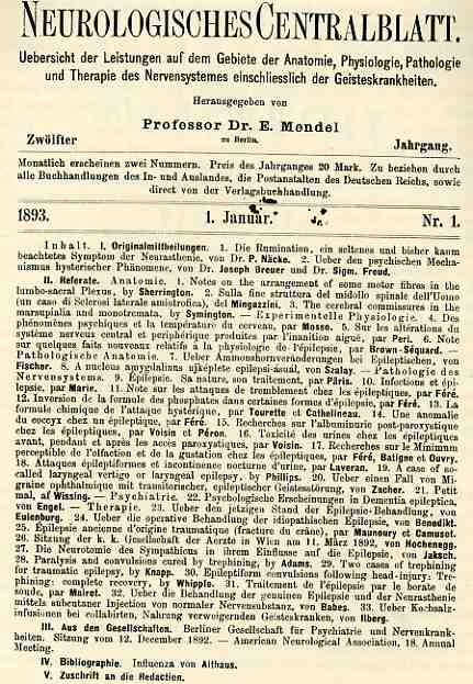
    Hidden in plain but complex view of this title page for the Neurologisches Centralblatt is  an early and important work by Sigmund Freud and Josef Breuer, “Ueber den psychischen Mechanismus hysterischer Phaenomen”.  Its a paper that establishes a Freudian dictum–based in the well-boiled ideas and work of Charcot–stating that it is not  mental trauma that leads to the cause of hysterical symptoms, but the memory of it. “Hysterical patients suffer mainly from reminiscences.” Ernst Jones explains:  “The trauma is not an evoking or precipitating factor, but rather, in its memory trace…resembles a foreign body that continues to irritate the mind”.

    Honestly I don’t know why people think about Freud.

    If for no other reason, this paper was important as it lead to a much more significant paper in Freud’s development of himself, Studien ueber Hysteria (1895). 

    The attraction for me was the complex presentation of the contents of this monthly journal–there’s a lot of data presented, it is not terribly reader-friendly, but it is still, well, pretty.  To me, its art.  Freud just  kinda mucks it up. 


  • A Woman Doctor Finds Her Name in Spite of it All, 1695

    JF Ptak Science Books LLC   Post 422

    0--ebay-dec 10 midwife234
    “Good wives and good plantations are the products of a good husband.”
    –Benjamin Franklin 1736

    “The best furniture in a house is an obedient wife”  Thomas 1808

    “A little house well filled, a little land well tilled, and a little wife well willed, are great riches ” John Ray 1683  [Most tidy this up by leaving off the last third of this famous little saying.]

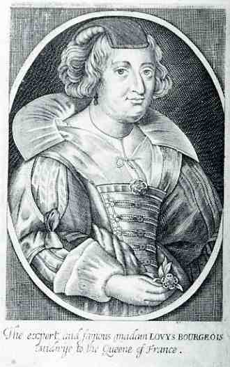
    The four anonymous/initialed authors on the title page of the first edition of The Complete Midwwife’s Practice (1659)…..claimed this book to be the first and best of its kind “since the birth of mankind” (!, chapter 4). That said, much of it was cribbed from the work of Louyse Bourgois (1564-1636), who had published  (though not under her own name) her obstetric secrets earlier in the century.   Bourgois was yet another in a long line of women who contributed to this science but whose identities were largely and obediently left to pervious memory among male authors—except in this case, where, in the fourth edition of this book, Bourgois was included in the longish-titled title page and actually has her engraved portrait included in the book.  And thus some small level of payback it realized.

    Recognizing the female’s importance in the writing of this book or not, the text of The Complete Midwife is still strongly constructiviist in maintaining the woman’s lesser role in most aspects of reproduction, as well as many aspects outside the biological realm.

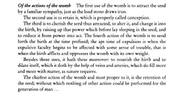
    For example, we find this state-of-the-art nugget:

    Blog--midwife

     This of course was nothing to raise an eyebrow over in the late 17th century–it was the state of medical knowledge seasoned with strong doses of female subjugation and slavery.

    The authors of this book were revealed by the fourth edition, and one of them comes from the long line of highly questionable obstetricians and medical monsters, the Chamberlain family.
    (I touched on them briefly here.) The family invented the forceps and kept the invention–which although simple was a huge aid in childbirth–secret, a utility controlled by them alone, completely in the dark, for more than 100 years.  Their secretivenesses protected the family fortune and future at a cost of untold numbers of difficult deliveries and, I’m sure, 100,000+ women and children lost in childbirth difficulties who could’ve been saved otherwise had the forceps been publicized and shared.  But this was not their lot, and nt their responsibility, nor their evident moral obligation…

    PreformationBut this jealous protection of their intellectual rights seeped out
    in other ways too, as in this very book, where this Chamerlain
    complained against one of his very own co-authors (the famous Nicholas
    Culpepper), who adopted a more popular approach to medicine, arguing
    for printing medical texts int he vernacular.  Perhaps this failing was
    genetic.

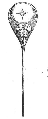
    Perhaps the worst of the panoply of wide ideas biologically inferiorizing (?) women belongs to  Nicolas Hartsoeker  (1626-1683) who discovered “animaclues” in sperm:  whole, completely formed, “little men”, who would be implanted into the furnace of woman to cook and nature them until they could be born.  This basically reduced woman to a piece of furniture–an oven–with all the great necessaries of reproduction performed by the man, a creation called the Homonculus completely generated by them,  with woman needed only as the fertile soil to plant the whole thing in.  

    ·Posted by :

    ·in:


  • Manly Purity & Beauty; Soap vs. The Royal Evil, 1889

    JF Ptak Sceince Books  LLC  Post 400

    0--blog---Nov 28 crimson chin146

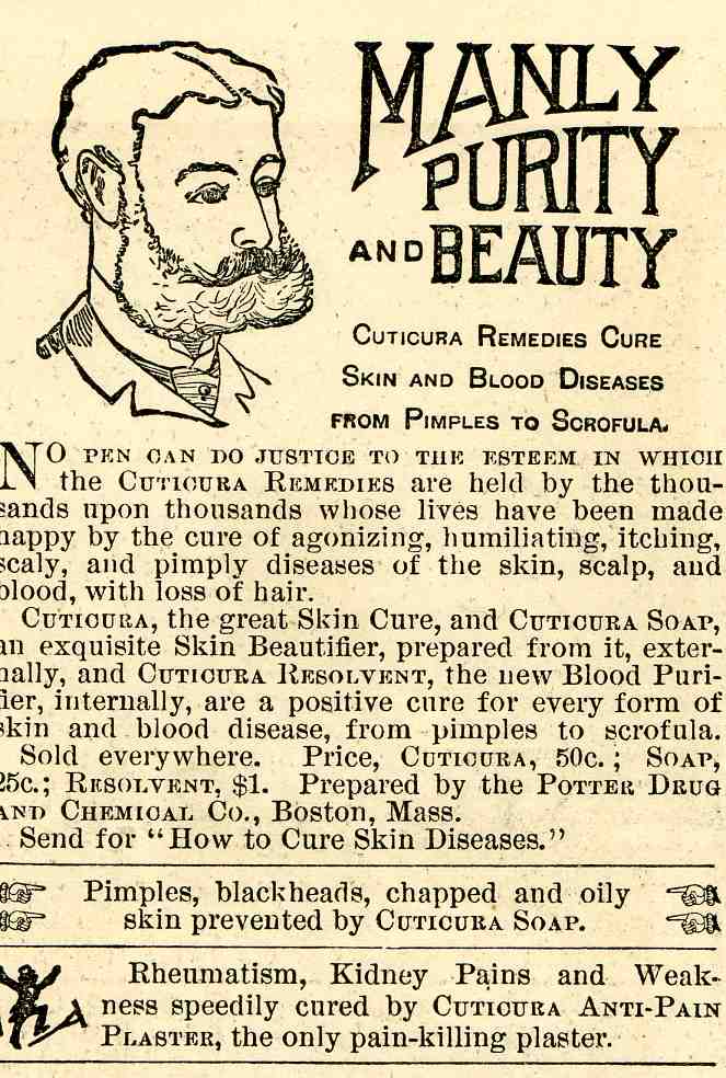
    This is 1889’s mini-version of a gorgeous male, a man of purity, virility, and pimple-free skin.  Also of scrofula-free skin, a condition description that seems to have faded from use after wide popularity for 500 years.  The picture of this man was used by the Cuticura Soap Company (actually the Potter Chemical and Drug Company of Boston) to promote its product, a soap with a bit of prussian blue tincture and some other stuff whose manufacturers claimed would (not could) cure all manner skin aliments, including the loss of hair.  The cure of scrofula–a form of tuberculosis affecting the lymph nodes of the neck and appearing as large welt-like dry abscesses–was no minor claim.  Aside from its quack connection, the claim was also an affront to royalty, as for hundreds of years the skin condition of scrofula (“the Royal Evil”)  was thought to be curable by a monarch’s touch; Henry IV was said to have touched thousands of people in this way.   There was also a religious connection to the sovereign touch cure, with a ceremony included in the Book of Common Prayer from the Anglican Church.  It survived in England until George I found the practice offensive and “too Catholic”. 

    An offshoot, Cuticura Resolvent, was offered to cure “every sort of blood disease”, while yet another product, Cuticura Anti-Pain Plaster, would cure rheumatism and kidney “pains and weaknesses”.  That’s quite a heap of expectation from soap, and the American Medical Association too it to task as early as 1912, calling the soap’s effectiveness as a curative “remote”; somewhat earlier, in 1908, the British Medical Association found the use of Cuticura for its stated curative capacities for syphilis to be “without basis”.  Pretty benign and reserved, I’d say, compared to what the AMA and BMA had to say about similar products. 

    The Cuticura identity is still in use–and popular–today, 143 years old, though so far as I know the modern makers make no claims for its soap to cure the Royal Evil.  Today its just soap.

    Crimson chin

    I’ll take this opportunity to reference this ideal of Manliness and Purity to a certain character from my younger daughter Tessie’s cartoon program, The Fairly Oddparents--this old man bears some resemblance to one Mr. Crimson Chin from that show.  (I just like referencing kids’ shows.)

    ·Posted by :

    ·in:

    —

  • “Correcting” Sight; Vision and Seeing

    JF Ptak Science Books LLC  Post 397

    0-blog---Nov 17 eyes078 

    Georg Bartisch (1535-1606) a brought-up-from-norhing-couldn’t-write-but-could-read barber-surgeon, from Dreseden, provided this spectacular illustration for his Ophthalmodouleia Das ist Augendienst (1583) (Treatment of the Eye).  As opportunistic as this image appears for imaginative interpretation, it was purely a cloth over the eyes to “help” correct strabismus (crossed eyes).   The slits in the cloth were meant to force the muscles of the eye to conform to their normal positions.

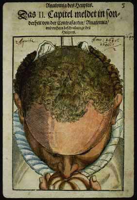
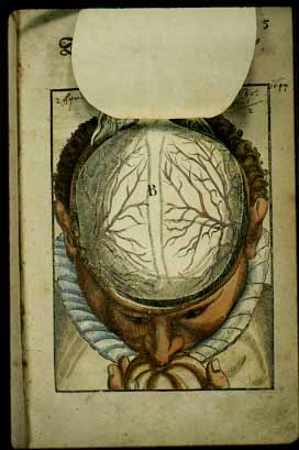
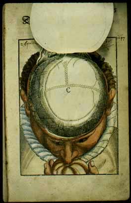
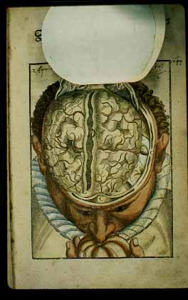
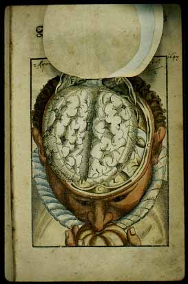
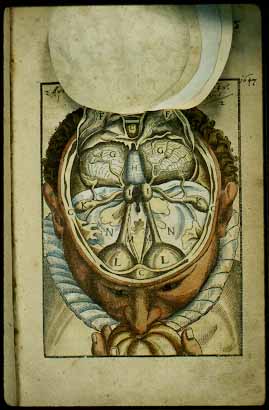
    More substantial, and more magnificent, are a series of anatomical dissections, flap-books if you will of superimposed moveable sheets of paper, showing different levels of the human body.  Finding these in 16th century books are quite rare, and I’ve included a series of six images showing the cut-aways of a dissection through the top of the head.  It is fabulous, and accurate.

    The images are all courtesy of :

    Duke University Medical Center Library
    History of Medicine Collections, Durham, NC
    Photographed by Dr. George O.D. Rosenwasser

    Flaps1

    Flaps2

    Flaps3

    Flaps4

    Flaps5


    Flaps6

    ·Posted by :

    ·in: